细胞-ID® 红色长期细胞示踪试剂盒(流式细胞仪和荧光显微镜) Cyto-ID® Red long-term cell tracer kit for flow cytometry and fluorescence microscopy

细胞-ID® 红色长期细胞示踪试剂盒(流式细胞仪和荧光显微镜)
Cyto-ID® Red long-term cell tracer kit for flow cytometry and fluorescence microscopy

  • 产品特性
  • 相关资料
  • Q&A
  • 参考文献

细胞示踪试剂盒细胞-ID®  红色长期细胞示踪试剂盒(流式细胞仪和荧光显微镜)                              Cyto-ID®  Red long-term cell tracer kit for flow cytometry and fluorescence microscopy


产品编号

产品名称

规格

ENZ-51036-K025

Cyto-ID Green lone-term cell tracer kit for flow cytometry and microscopy

25 assays

ENZ-51037-K025

Cyto-ID Red long-term cell tracer kit for flow cytometry and fluorescence microscopy

25 assays

相关产品资料请点击下载:活细胞荧光分析 Ver.3


※ 本页面产品仅供研究用。研究以外不可使用。


产品列表
产品编号 产品名称 产品规格 产品等级 备注
ENZ-51036-K025 Cyto-ID®  Green long-term cell tracer kit for flow cytometry and microscopy 
细胞-ID ®  绿色 长期细胞示踪试剂盒 (流式细胞仪和荧光显微镜)
25 assays
ENZ-51037-K025 Cyto-ID®  Red long-term cell tracer kit for flow cytometry and fluorescence microscopy 
细胞-ID ®  红色 长期细胞示踪试剂盒 (流式细胞仪和荧光显微镜)
25 assays
免责声明

1. 本公司密切关注本网站发布的内容,但不保证发布内容的准确性、完整性、可靠性和最新性等。

2. 本公司不保证使用本网站期间不会出现故障或计算机病毒污染的风险。

3. 无论何种原因,使用本网站时给用户或第三方造成的任何不利或损害,本公司概不负责。此外,对于用户与其他用户或第三方之间因本网站发生的任何交易、通讯

3. 纠纷,本公司概不负责。

4. 本网站可提供的所有产品和服务均不得用于人体或动物的临床诊断或治疗,仅可用于科研等非医疗目的。如任何用户将本网站提供的产品和服务用临床诊断或治

4. 疗,以及他特定的用途或行为,本公司概不保证其安全性和有效性,并且不负任何相关的法律责任。

CYTO-ID® 自噬检测试剂盒 2.0 CYTO-ID® Autophagy detection kit 2.0

CYTO-ID® 自噬检测试剂盒 2.0
CYTO-ID® Autophagy detection kit 2.0

  • 产品特性
  • 相关资料
  • Q&A
  • 参考文献

CYTO-ID® 自噬检测试剂盒 2.0CYTO-ID® 自噬检测试剂盒 2.0                              CYTO-ID® Autophagy detection kit 2.0

  CYTO-ID® Autophagy Detection Kit 2.0 使用新型染料检测自噬小泡和监控活细胞自噬流,选择性标记积累的自噬小泡。染料已通过优化,不染溶酶体,在自噬前体、自噬体和自噬溶酶体里呈现出明 亮的荧光。该试剂盒提供了一种无需细胞转染,可以在活细胞中监控细胞自噬的快速定量方法。

◆特点


  ● 更明亮,更耐光,特异性染色自噬小泡

  ● 无需转染及转染效率验证

  ● 快速量化原生异质性细胞群中的自噬

  ● 不染溶酶体,减少其他染料的背景干扰

   便于高通量筛选自噬激活剂和抑制剂


◆原理


  该产品所用探针是阳离子两亲性示踪剂(CAT)染料,以类似诱导磷脂药物的方式迅速进入到细胞中。染料的功能基团能够选择性标记与自噬通路相关的小泡,而不会在溶酶体中聚集。


胞内物质被扩大的膜囊包裹,吞噬泡形成双层膜囊泡,成为自噬体。自噬体外膜随后与溶酶体融合,和内部物质被自噬性溶酶体降解。自噬的各种调节因子也被描绘于图中。

CYTO-ID® 自噬检测试剂盒 2.0                              CYTO-ID® Autophagy detection kit 2.0

自噬原理图

◆应用


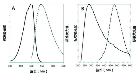
CYTO-ID® 绿色检测试剂2(A组)在PBS确定的吸光度和荧光图谱(499/548 nm)。


Hoechst33342(图B)在1X测定缓冲液来确定的吸光度和荧光图谱(350 /461 nm)。

的发放

CYTO-ID® 自噬检测试剂盒 2.0                              CYTO-ID® Autophagy detection kit 2.0

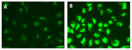
HeLa cells使用 CYTO-ID® Green Detection Reagent 2进行染色


(A) 完全培养基 (B) 在饥饿培养基 (EBSS)中添加40 µM Chloroquine培养4 h 。在含有Chloroquine 的EBSS培养基中培养的细胞表现非常明亮的绿色荧光信号和点状结构。

CYTO-ID® 自噬检测试剂盒 2.0                              CYTO-ID® Autophagy detection kit 2.0


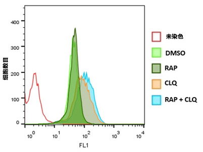
运用流式细胞术分析Jurkat细胞的自噬


通过0.5 µM Rapamycin (RAP), 10 µM Chloroquine (CLQ)处理或不处理Jurkat细胞,或两者一起处理20 h。CYTO-ID® Green Detection Reagent 2染色30 min后,洗脱,用流式细胞仪分析。直方图叠加呈现结果。用RAP+CLQ处理细胞显示荧光有升高。

CYTO-ID® 自噬检测试剂盒 2.0                              CYTO-ID® Autophagy detection kit 2.0

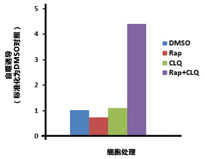
微孔板分析HepG2细胞的自噬


HepG2经过DMSO(对照),0.5 μM Rapamycin (Rap), 10 µM Chloroquine (CLQ),或0.5 µM Rap 和10 µM CLQ同时培养20 h。细胞经过Hoechst 33342染色进行细胞数目标准化。Rap 和CLQ同时处理细胞显示自噬作用上升。

CYTO-ID® 自噬检测试剂盒 2.0                              CYTO-ID® Autophagy detection kit 2.0

◆相关产品


产品编号

产品名称

规格

备注

应用

ENZ-51002-25

GFP-Certified® Apoptosis/Necrosis detection kit

细胞凋亡/坏死检测试剂盒

25 assays

多重检测,区分正常、

早期凋亡、晚期凋亡和坏死细胞,与GFP和其他绿色荧光探针兼容。

FC,荧光显微镜,荧光检测

ENZ-51002-100

100 assays

ENZ-51021-K200

Nuclear-ID® Green

hromatin condensation detection kit

细胞核绿色染色体皱缩检测试剂盒

1 Kit

高渗透性的绿色荧光染色增强了细胞凋亡诱导染色质固缩。

FC,荧光显微镜,荧光检测

ENZ-52406

NUCLEAR-ID® Red DNA stain

DNA染色试剂盒(红色荧光)

200 µL

细胞可渗透的DNA染色应用广泛。

≥93%(HPLC),FC,荧光检测

ENZ-CHM103-0200

Nuclear-ID® Blue

DNA stain   (GFP-Certified®)

Nuclear ID® 蓝色DNA染色(GFP细胞系)

200 µL

细胞可渗透的DNA染色应用广泛。

≥93%(HPLC),FC,荧光检测

ENZ-51015-KP002

Lyso-ID® Red

cytotoxicity kit (GFP-Certified®)

溶酶体细胞毒理检测试剂盒(红色荧光)(绿色细胞系)

1 Kit

快速,定量和HTS-兼容的检测活细胞毒性试剂。

荧光显微镜,荧光检测

ENZ-51035-0025

PROTEOSTAT® Aggresome etection kit
蛋白内稳态® 聚集体 检测试剂盒

25 tests

Robust、定量的聚集小体用于神经退行性疾病,肝病和毒理学研究

FC,荧光显微镜,荧光检测

ENZ-51035-K100

100 tests

相关产品资料请点击下载:活细胞荧光分析 Ver.3

自噬技术文章:


通过流式细胞术定量自噬解释与白血病肿瘤生长的联系

Posted By Morgan Mathieu

Tags: Autophagy, Cancer


主要参考文献


(1)


A live-cell fluorescence microplate assay suitable for monitoring vacuolation arising from drug or toxic agent treatment: J. Coleman, et al.; J. Biomol. Screen. 15, 398 (2010), 摘要;

(2)


Methods in mammalian autophagy research: N. Mizushima, et al.; Cell 140, 313 (2010),摘要;

(3)


Assays to Assess Autophagy Induction and Fusion of Autophagic Vacuoles with a Degradative Compartment, Using Monodansylcadaverine (MDC) and DQ-BSA: C.L. Vazquez & M.I. Colombo; Methods Enzymol. 452, 85 (2009),摘要;

(4)


Desmethylclomipramine induces the accumulation of autophagy markers by blocking autophagic flux: M. Rossi, et al.; J. Cell Sci. 122, 3330 (2009),  摘要;

(5)


Guidelines for the use and interpretation of assays for monitoring autophagy in higher eukaryotes: D.J. Klionsky, et al.; Autophagy 4, 151 (2008),  摘要;

产品列表
产品编号 产品名称 产品规格 产品等级 备注
ENZ-KIT175-0200 Cyto-ID® Autophagy detection kit 2.0
 CYTO-ID®自噬检测试剂盒2.0
200 tests
免责声明

1. 本公司密切关注本网站发布的内容,但不保证发布内容的准确性、完整性、可靠性和最新性等。

2. 本公司不保证使用本网站期间不会出现故障或计算机病毒污染的风险。

3. 无论何种原因,使用本网站时给用户或第三方造成的任何不利或损害,本公司概不负责。此外,对于用户与其他用户或第三方之间因本网站发生的任何交易、通讯

3. 纠纷,本公司概不负责。

4. 本网站可提供的所有产品和服务均不得用于人体或动物的临床诊断或治疗,仅可用于科研等非医疗目的。如任何用户将本网站提供的产品和服务用临床诊断或治

4. 疗,以及他特定的用途或行为,本公司概不保证其安全性和有效性,并且不负任何相关的法律责任。

CYTO-ID® 自噬检测试剂盒 CYTO-ID® Autophagy detection kit

CYTO-ID® 自噬检测试剂盒
CYTO-ID® Autophagy detection kit

  • 产品特性
  • 相关资料
  • Q&A
  • 参考文献

CYTO-ID® 自噬检测试剂盒CYTO-ID® 自噬检测试剂盒                              CYTO-ID® Autophagy detection kit

  CYTO-ID® Autophagy Detection Kit 使用新型染料检测自噬小泡和监控活细胞自噬流,选择性标记积累的自噬小泡。染料已通过优化,不染溶酶体,在自噬前体、自噬体和自噬溶酶体里呈现出明亮的荧光。该试剂盒提供了一种无需细胞转染,可以在活细胞中监控细胞自噬的快速定量方法。

CYTO-ID® 自噬检测试剂盒                              CYTO-ID® Autophagy detection kit

CYTO-ID® Autophagy Detection Kit

◆特点

● 无需转染,定量监测活细胞中的自噬情况

● 免去 LC3-GFP 转染所需的时间和精力以及转染效率的验证

● 含有特殊基团的专利染料选择性地染色自噬小泡

● 试剂盒中包含已知活性的自噬抑制剂与激活剂

● 快速量化原生异质性细胞群中的自噬

● 选择性地全方位染色,可检测和区分自噬流(autophagic flux)和自噬溶酶体的积累

● 不染溶酶体,减少其他染料的背景干扰

 便于高通量筛选自噬激活剂和抑制剂

◆原理


  该产品所用探针是阳离子两亲性示踪剂(CAT)染料,以类似诱导磷脂药物的方式迅速进入到细胞中。染料的功能基团能够选择性标记与自噬通路相关的小泡,而不会在溶酶体中聚集。


胞内物质被扩大的膜囊包裹,吞噬泡形成双层膜囊泡,成为自噬体。自噬体外膜随后与溶酶体融合,和内部物质被自噬性溶酶体降解。自噬的各种调节因子也被描绘于图中。

CYTO-ID® 自噬检测试剂盒                              CYTO-ID® Autophagy detection kit

自噬原理图

◆应用


省时省力,无需转染即可自快速全面的标记自噬小泡


为了演示 CYTO-ID® Green detection reagent 的优势,先用 RFP-LC3 表达载体转染 HeLa 细胞,10 μM 的 Tamoxifen 处理过夜,然后用 CYTO-ID® 绿色检测试剂染色。LC3 转染法需要过夜,而 CYTO-ID® 绿色检测试剂的方法在15-30分钟内标记细胞达100%。图A:绿信号显示自噬小泡的 CYTO-ID® 绿染;图B:成功转染细胞的 RFP-LC3 表达(红色);图C:自噬体的特异性标记 LC3,与 CYTO-ID® 绿色染料标记的小泡共定位的复合图像。

的发放

CYTO-ID® 自噬检测试剂盒                              CYTO-ID® Autophagy detection kit


无转染的自噬检测


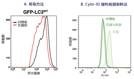
Hela细胞进行饥饿与恢复,然后使用 CYTO-ID® Green detection reagent 标记。染料可以明确检测和量化自噬及与自噬诱导相关的自噬小泡前体。图1A:稳定表达 GFP-LC3 的CHO细胞,结果显示对照组与饥饿的细胞群的相对较差的基线分离,自噬难以量化。图1B:CYTO-ID® Autophagy Detection Kit 特异性标记 LC3 蛋白依赖的自噬小泡,免去转染操作。

CYTO-ID® 自噬检测试剂盒                              CYTO-ID® Autophagy detection kit


自噬积累和自噬流的可视化


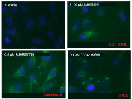
可用荧光显微镜观察CYTO-ID® 自噬绿色染料检测的自噬液泡积累和自噬流。Hela细胞用以下试剂进行模拟诱导,0.2% DMSO(A)和100 μM  Clonidine hydrochloride(B)进行诱导,5 μM Loperamide hydrochloride 和 1 μM PP242 hydrate(D)在37°C诱导12小时。处理后,细胞在37℃下用CYTO-ID® Green Detection reagent 孵育10分钟,再用分析缓冲液清洗。细胞核被 Hoechst 33342 染料复染为蓝色。

CYTO-ID® 自噬检测试剂盒                              CYTO-ID® Autophagy detection kit


避免非特异性溶酶体染色的背景干扰


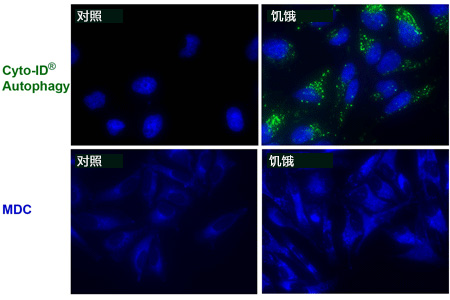
相比其他 MDC 染色溶酶体检测,CYTO-ID® 绿色染料无溶酶体染色背景,CYTO-ID® Autophagy kit 无需紫外活细胞分析,在显微镜共标记应用中,与 Hoechst 染料兼容。

CYTO-ID® 自噬检测试剂盒                              CYTO-ID® Autophagy detection kit


使用流式细胞仪通过 CYTO-ID® Autophagy Detection 

Kit 分析自噬


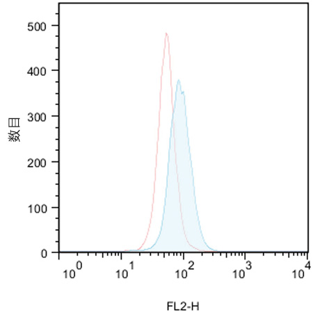
对照组(红线峰值)无诱导,10 uM Tamoxifen(ALX-550-095)处理 Jurkat 细胞(白血病T细胞)(蓝线峰值)。处理18小时后,细胞结合 CYTO-ID® 绿色检测试剂,然后不经过流式细胞仪清洗,进行分析。结果通过直方图呈现。对照组细胞被染色,但荧光亮度低。实验组中,CYTO-ID® 绿色荧光信号增加约2倍,表明 Tamoxifen 处理能引起 Jurkat 细胞中自噬的增加。


CYTO-ID® 自噬检测试剂盒                              CYTO-ID® Autophagy detection kit

CYTO-ID® 自噬检测试剂盒                              CYTO-ID® Autophagy detection kit

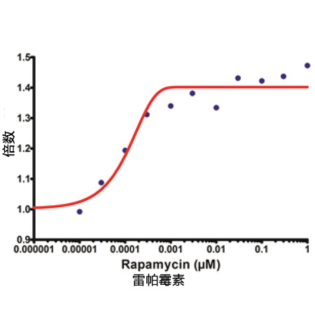
使用 mTOR 激酶抑制剂 Rapamycin 处理 HepG2 细胞孵育过夜,结果显示 CYTO-ID® 染色信号升高。

CYTO-ID® 自噬检测试剂盒                              CYTO-ID® Autophagy detection kit

CYTO-ID® 绿色染料大多与 RFP-LC3蛋白共定位。0.1 μM Rapamycin(典型自噬诱导剂)处理过夜,转染 Hela 细胞表达RFP-LC3。

A: CYTO-ID® Green 染色; B: RFP-LC3; C: 合成图像。

CYTO-ID® 自噬检测试剂盒 2.0

◆相关产品

产品编号

产品名称

规格

备注

应用

ENZ-51002-25

GFP-Certified®Apoptosis/Necrosis detection kit

细胞凋亡/坏死检测试剂盒

25 assays

多重检测,区分正常、

早期凋亡、晚期凋亡和坏死细胞,与GFP和其他绿色荧光探针兼容。

FC,荧光显微镜,荧光检测

ENZ-51002-100

100 assays

ENZ-51021-K200

Nuclear-ID®Green

hromatin condensation detection kit

细胞核绿色染色体皱缩检测试剂盒


1 Kit

高渗透性的绿色荧光染色增强了细胞凋亡诱导染色质固缩。


FC,荧光显微镜,荧光检测

ENZ-52406

NUCLEAR-ID®Red DNA stain

DNA染色试剂盒(红色荧光)

200 µL

细胞可渗透的DNA染色应用广泛。

≥93%(HPLC),FC,荧光检测

ENZ-CHM103-0200

Nuclear-ID® Blue

DNA stain   (GFP-Certified®)

Nuclear ID® 蓝色DNA染色(GFP细胞系)

200 µL

细胞可渗透的DNA染色应用广泛。

≥93%(HPLC),FC,荧光检测

ENZ-51015-KP002

Lyso-ID® Red

cytotoxicity kit   (GFP-Certified®)

溶酶体细胞毒理检测试剂盒(红色荧光)(绿色细胞系)

1 Kit

快速,定量和HTS-兼容的检测活细胞毒性试剂。

荧光显微镜,荧光检测

ENZ-51035-0025

PROTEOSTAT®Aggresome   etection kit

蛋白内稳态聚集体 检测 试剂盒

25 tests

Robust、定量的聚集小体用于神经退行性疾病,肝病和毒理学研究

FC,荧光显微镜,荧光检测

ENZ-51035-K100

100 tests


处理

目的

效果

μM

诱导时间 (hrs)

细胞系

饥饿

抑制哺乳动物Rapamycin (mTOR)

激活自噬

N/A

1~4

HeLa, HepG2, Jurkat

Rapamycin

抑制哺乳动物Rapamycin (mTOR)

激活自噬

0.2

6~18

HeLa, Jurkat

PP242

mTOR ATP-竞争性抑制剂

激活自噬

1

18

HeLa

Lithium

抑制IMPase 和降低inositol 和IP3水平; mTOR依赖性

激活自噬

10,000

18

HeLa, Jurkat

Trehalose

未知, mTOR依赖性

激活自噬

50,000

6

HeLa, Jurkat

Bafilomycin A1

抑制 Vacuolar-ATPase

抑制自噬

6~9*10-3

18

HeLa, Jurkat

Chloroquine

碱化 Lysosomal pH

抑制自噬

10~50

18

HeLa, Jurkat

Tamoxifen

增加细胞内 ceramide的水平和消除PI3K的抑制效果

激活自噬

4~10

6~18

HeLa, HepG2, Jurkat

Verapamil

钙离子通道阻滞剂;降低胞浆内Ca2+水平; mTOR依赖性

激活自噬

40~100

18

HeLa, Jurkat

HydroxyChloroquine

Alkalinizes Lysosomal pH

抑制自噬 

10

18

HeLa, Jurkat

Loperamide

Ca2+ 通道阻滞剂;降低胞浆内 Ca2+浓度; mTOR依赖性

激活自噬

5

18

HeLa

Clonidine

咪唑啉-1受体激动剂; 降低 cAMP 水平; mTOR依赖性

激活自噬

100

18

HeLa

MG-132

选择性蛋白酶抑制剂

激活自噬

2~5

18

HeLa, Jurkat

Norclomipramine

碱化Lysosomal pH

抑制自噬

5-20

18

HeLa

Epoxomicin

选择性蛋白酶抑制剂

诱导聚集体

0.5

18

HeLa

Velcade®

选择性蛋白酶抑制剂

诱导聚集体

0.5

18

HeLa

Amyloid beta peptide 1-42

引起氧化应激

诱导聚集体

25

18

SK-N-SH

Cyto-ID®检测试剂盒 Q&A


Q:Cyto-ID®自噬检测试剂与活细胞孵育的最长时间是多少(几分钟/几小时/几天)?
A:我们建议 37℃ 孵育不超过 30 分钟。很多实验表明,孵育 1 小时不会伤害细胞。孵育超过 1 小时,一些细胞看上去状态不好。


Q:是否可用荧光显微镜观察 3D 培养细胞的自噬?
A:我们没有用该染料染过 3D 细胞,但是一小撮细胞是可以染色的。活体组织的话,由于染料的渗透性不好,不能进入样本的内部,所以效果不

A:想。染白细胞是可以。


Q:用双重染料染在 37℃ 孵育 30 分钟后,细胞状态不好开始皱缩。有些细胞也从板上脱离,阳性对照和 DMSO 对照没有观察到特异性染色。
A:对于比较敏感的细胞,优化流程如下:
A:1. 使用 PBS/5% FBS 或者完全培养基作为 assay buffer,最好不含酚红。
A:2. 如果细胞状态仍然不好,建议染色时间缩短至 15~20 分钟。
A:3. 减少染料的浓度至 1000× 稀释。需要显微镜观察时延长荧光照射的时间。
A:4. 可能光褪色会导致信号模糊。建议使用封片剂防止光褪色。


Q:用流式检测的话,样品在用 Cyto-ID®自噬检测试剂孵育后多久可用流式观察?操作手册是说 30 分钟。是否可以在检测之前短暂的将样品放

A:在冰上?
A:可以将样品放在冰上延长孵育的时间。一般的经验法则,尽可能缩短活细胞与染料的结合。缺氧,低温或者其他应激状态也可以诱导自噬。


Q:GFP-CertifiedTM 细胞凋亡/坏死检测试剂盒(ENZ-51002),Cyto-ID® 自噬检测试剂盒(ENZ-51031)是否可以同时染色,检测细胞凋

A:亡、坏死和自噬?
A:GFP-CertifiedTM 细胞凋亡/坏死检测试剂盒(ENZ-51002)和 Cyto-ID® 检测试剂盒(ENZ-51031)不能配合使用,因为细胞凋亡和自噬检

A:测的染料都发绿色荧光。这两个试剂盒可以平行使用。比如,用十字孢碱(Staurosporine)处理细胞,一部分样品用 GFP-CertifiedTM 细胞

A:凋亡/坏死检测试剂盒检测,一部分样品用 Cyto-ID® 检测试剂盒检测。


Q:Cyto-ID®自噬检测试剂盒(ENZ-51031)的 Cyto-ID®自噬检测试剂能否与 ProteoStat®蛋白聚集检测(ENZ-51023)的 ProteoStat®

A:检测试剂联合使用?
A:Cyto-ID®绿色自噬检测试剂与 ProteoStat®检测试剂发的荧光颜色不同,他们可用于同一实验,客户需要优化实验流程。

Q:Cyto-ID®自噬检测试剂的原理是什么?
A:染料是申请专利的。该染料是阳离子两性示踪剂(CAT)染料,跟很多阳离子化合物一样可以快速进入细胞。染料穿过细胞膜双分子层后被动

A:扩散,不需要蛋白结合或者转运子活性。可滴定部分的筛选使的染料不会再溶酶体聚集,能够标记自噬通道相关的液泡。另外,染料的发射光

A:密度增强后可以标记自噬液泡相关片层膜结构。我们可以提供染料染自噬液泡的相关数据。诱导自噬溶酶体,比如用氯喹或者baflimyocin 处

A:理后,会导致荧光变亮(250~300%)。雷帕霉素诱导自噬后能够用3-MA 抑制。在EBSS 中氨基酸缺乏会导致1 小时后在自噬液泡中染料的

A:积累,这种积累是可逆的(补充营养后即可恢复)。我们有一系列用于研究自噬的化合物(Screen-Well®Autophagy library)


Q:与单丹磺酰尸胺(MDC)相比,Cyto-ID®自噬检测试剂有哪些优势?
A:MDC 需要 365 nm UV 荧光照明,与通常流式配置的 488 nm 激发源不兼容。Cyto-ID®自噬染料是 488 nm 激发波长,绿色的发射荧光,可

A:以高亮自噬通道不同的液泡成分。需要注意的是,与 lysomotrophic 染料不同,MDC、LysoTrackerTMRed、吖啶橙主要是染溶酶体的,

A:Cyto-ID®自噬染料只能染一点点溶酶体,主要是染自噬溶酶体和早期自噬部分的选择标记物。


Q:与 GFP-LC3 转染细胞相比,Cyto-ID®自噬检测试剂盒(ENZ-51031)的优势是什么?
A:Cyto-ID®检测试剂盒(ENZ-51031)的优势在于不用转染细胞。转染很短暂,不同实验需要再购买相关试剂。由于细胞过表达,会导致

A:GFP-LC3 聚集,结果呈假阳性。转染效率低于100%,有些细胞不会表达必要的 GFP 组件。该试剂盒检测的自噬,细胞群的一致性会更好。

A:不需要每个细胞系都进行转染。也可以标记原代细胞。原代细胞不容易进行转染。


Q:有哪些细胞系或者原代细胞可用 Cyto-ID®自噬检测试剂?
A:用该试剂盒见检测过的代表性的细胞和细胞系包括肝细胞、SK-N-SH 神经瘤细胞、CHO 细胞、骨肉瘤来源细胞、黑素留细胞、乳腺癌细胞、

A:宫颈癌细胞、卵巢癌细胞、B 淋巴瘤细胞、结肠癌细胞、HepG 细胞、Jurkat T细胞、牛大动脉内皮细胞(BAEC)。


Q:Cyto-ID®自噬检测试剂能否与其他染料配合使用,区分细胞核内体和自噬内涵体?
A:Cyto-ID®红色长期示踪剂染料(ENZ-51037)能够染细胞质膜,通过细胞内吞存在于细胞中。Cyto-ID®红色染料与 Cyto-ID®自噬检测试剂

A:能够高亮异体吞噬 vs 自噬。也可以 Cyto-ID®自噬检测试剂与Lyso-ID®红色染料或者LysoTrackerTM 红色染料进行活细胞染色,荧光显微镜

A:观察。


Q:Cyto-ID®自噬检测试剂的稳定性如何?
A:荧光染料会发生光褪色,但是这种染料比荧光素更稳定。按照操作手册出来荧光染料。

Q:Cyto-ID®自噬检测试剂盒染色后是否可固定细胞?
A:在操作手册中提供了固定细胞的流程。不推荐用表面活性剂给细胞打孔,定位的荧光染料丢失。


Q:活细胞检测时,荧光信号强度可维持多长时间?
A:细胞诱导后发生自噬,在分析前染色。诱导时间少于 1 小时或者诱导几天,诱导时间取决于自噬诱导剂。我们没有在活细胞上进行长时间的染

A:色,有客户发现可以至少染色 24 小时。


参考文献


1.

A comparison of strategies for immortalizing mouse embryonic fibroblasts: M.M. St. Amad, et al.; J. Biol. Methods 3, e41 (2016), Application(s): Detected autophagy induction in SV40 transformed versus serially passed MEFs, 全文

2.

Astemizole-Histamine induces Beclin-1-independent autophagy by targeting p53-dependent crosstalk between autophagy and apoptosis: R. Jakhar, et al.; Cancer Lett. 372, 89 (2016), Application(s): Flow cytometry analysis, 摘要;

3.

Atg5 Is Essential for the Development and Survival of Innate Lymphocytes: T.E. O'Sullivan, et al.; Cell Rep. 15, 1910 (2016), Application(s): Liver lymphocytes were harvested and stained with cell surface antibodies and then incubated with 1:400 Cyto-ID Autophagy Detection Reagent, 摘要全文

4.

Autophagy-related gene 5 and Wnt5 signaling pathway requires differentiation of embryonic stem cells into odontoblast-like cells: N. Ozeki, et al.; Exp. Cell Res. 341, 92 (2016), Application(s): Autophagy flux, 摘要;

5.

DHA-induced stress response in human colon cancer cells-focus on oxidative stress and autophagy: K. Pettersen, et al.; Free Radic. Biol. Med. 90, 158 (2016), Application(s):Autophagy determined by flow cytometry, 摘要;

6.

Diosgenin induces ROS-dependent autophagy and cytotoxicity via mTOR signaling pathway in chronic myeloid leukemia cells: S. Jiang, et al.; Phytomedicine 23, 243 (2016),Application(s): Confocal immunofluorescence, 摘要;

7.

Efavirenz causes oxidative stress, endoplasmic reticulum stress, and autophagy in endothelial cells: M. Weiss, et al.; Cardiovasc. Toxicol. 16, 90 (2016), 摘要;

8.

MiR-193b promotes autophagy and non-apoptotic cell death in oesophageal cancer cells: M.J. Nyhan, et al.; BMC Cancer 16, 101 (2016), Application(s): Assay used to stain live cells, 摘要全文

9.

(Pro)renin receptor regulates autophagy and apoptosis in podocytes exposed to high glucose: C. Li, et al.; Am. J. Physiol. Endocrinol. Metab. 309, E302 (2015), Application(s):Confocal microscopy using mouse podocytes, 摘要;

10.

A rapid and high content assay that measures cyto-ID-stained autophagic compartments and estimates autophagy flux with potential clinical applications: S. Guo, et al.; Autophagy11, 560 (2015), Application(s): Fluorescent detection, Microplate Reader , 摘要全文

11.

A Systems Approach Identifies Essential FOXO3 Functions at Key Steps of Terminal Erythropoiesis: R. Liang, et al.; PLoS Genet. 11, e1005526 (2015), Application(s):Autophagy flux measured by flow cytometry , 摘要全文

12.

ABT-888 enhances cytotoxic effects of temozolomide independent of MGMT status in serum free cultured glioma cells: R.K. Balvers, et al.; J. Transl. Med. 13, 74 (2015),Application(s): Assay, 摘要全文

13.

Activation of autophagy in response to nanosecond pulsed electric field exposure: J.C. Ullery, et al.; Biochem. Biophys. Res. Commun. 458, 411 (2015), Application(s):Fluorescence microscopy using U937 monocyte and CHO-K1 cell lines, 摘要;

14.

Aflatoxin biosynthesis is a novel source of reactive oxygen species-a potential redox signal to initiate resistance to oxidative stress?: L.V. Roze, et al.; Toxins (Basel). 7, 1411 (2015),Application(s): Assay, 摘要全文

15.

Alisertib induces cell cycle arrest and autophagy and suppresses epithelial-to-mesenchymal transition involving PI3K/Akt/mTOR and sirtuin 1-mediated signaling pathways in human pancreatic cancer cells: F. Wang, et al.; Drug Des. Devel. Ther. 9, 575 (2015), Application(s): Flow cytometry using PANC-1 and BxPC-3 pancreatic cancer cell lines, 摘要全文

16.

Alisertib, an Aurora kinase A inhibitor, induces apoptosis and autophagy but inhibits epithelial to mesenchymal transition in human epithelial ovarian cancer: Y.H. Ding, et al.; Drug Des. Devel. Ther. 9, 425 (2015), Application(s): Flow cytometry using SKOV3 and OVCAR-4 ovarian cancer cell lines, 摘要全文

17.

Andrographolide Analogue Induces Apoptosis and Autophagy Mediated Cell Death in U937 Cells by Inhibition of PI3K/Akt/mTOR Pathway: D. Kumar, et al.; PLoS One 10, e0139657 (2015), Application(s): Flow cytometric analysis of Cyto-ID Green Detection Reagent , 摘要全文

18.

Apoptotic Cell Death Induced by Resveratrol Is Partially Mediated by the Autophagy Pathway in Human Ovarian Cancer Cells: F. Lang, et al.; PLoS One 10, e0129196 (2015),Application(s): Live Cell Imaging, 摘要全文

19.

Araguspongine C induces autophagic death in breast cancer cells through suppression of c-Met and HER2 receptor tyrosine kinase signaling: M.R. Akl, et al.; Mar. Drugs 13, 288 (2015), Application(s): Flow cytometry using BT-474 breast cancer cell line, 摘要全文

20.

Autocrine VEGF maintains endothelial survival through regulation of metabolism and autophagy: C.K. Domigan, et al.; J. Cell. Sci. 128, 2236 (2015), 摘要;

21.

Autophagy is activated in systemic lupus erythematosus and required for plasmablast development: A.J. Clarke, et al.; Ann. Rheum. Dis. 74, 912 (2015), 摘要全文

22.

Autophagy limits proliferation and glycolytic metabolism in acute myeloid leukemia: A.S. Watson, et al.; Cell Death Discov. 1, 15008 (2015), Application(s): CytoID assay in human and mouse HSCs, 摘要全文

23.

Baicalin inhibits autophagy induced by influenza A virus H3N2: H.Y. Zhu, et al.; Antiviral Res. 113, 62 (2015), Application(s): Fluorescence microscopy using A549 human lung cancer cell line, 摘要;


24.

Bardoxolone methyl induces apoptosis and autophagy and inhibits epithelial-to-mesenchymal transition and stemness in esophageal squamous cancer cells: Y.Y. Wang, et al.; Drug Des. Devel. Ther. 9, 993 (2015), Application(s): Flow Cytometry, 摘要全文

25.

Cell-penetrating peptide derived from human eosinophil cationic protein inhibits mite allergen Der p 2 induced inflammasome activation: S.J. Yu, et al.; PLoS One 10, e0121393 (2015), Application(s): Flow cytometry of THP-1 leukemia cell line, 摘要全文

26.

Chemoproteomics Reveals Novel Protein and Lipid Kinase Targets of Clinical CDK4/6 Inhibitors in Lung Cancer: N.J. Sumi, et al.; ACS Chem. Biol. 10, 2680 (2015),Application(s): Quantification of autophagosomes, 摘要;

27.

Circulating hemocytes from larvae of the Japanese rhinoceros beetle Allomyrina dichotoma (Linnaeus) (Coleoptera: Scarabaeidae) and the cellular immune response to microorganisms: S. Hwang, et al.; PLoS One 10, e0128519 (2015), Application(s):Fluorescence microscopy using hemocytes from Japanese rhinoceros beetle Allomyrina dichotoma larvae, 摘要全文

28.

Citreoviridin induces ROS-dependent autophagic cell death in human liver HepG2 cells: Y.N. Liu, et al.; Toxicon. 95, 30 (2015), Application(s): Fluorescence microscopy using HepG2 cell line, 摘要;

29.

Clozapine induces autophagic cell death in non-small cell lung cancer cells: Y.C. Yin, et al.; Cell. Physiol. Biochem. 35, 945 (2015), 摘要;

30.

Coffee and caffeine potentiate the antiamyloidogenic activity of melatonin via inhibition of Aβ oligomerization and modulation of the Tau-mediated pathway in N2a/APP cells: L.F. Zhang, et al.; Drug Des. Devel. Ther. 9, 241 (2015), Application(s): Flow Cytometry,摘要全文

31.

Combination of the mTOR inhibitor RAD001 with temozolomide and radiation effectively inhibits the growth of glioblastoma cells in culture: H. Burckel, et al.; Oncol. Rep. 33, 471 (2015), 摘要;

32.

Danusertib Induces Apoptosis, Cell Cycle Arrest, and Autophagy but Inhibits Epithelial to Mesenchymal Transition Involving PI3K/Akt/mTOR Signaling Pathway in Human Ovarian Cancer Cells: D. Zi, et al.; Int. J. Mol. Sci. 16, 27228 (2015), Application(s): Confocal fluorescence microscopy, 摘要全文

33.

Danusertib, a potent pan-Aurora kinase and ABL kinase inhibitor, induces cell cycle arrest and programmed cell death and inhibits epithelial to mesenchymal transition involving the PI3K/Akt/mTOR-mediated signaling pathway in human gastric cancer AGS and NCI-N78 cells: C.X. Yuan, et al.; Drug Des. Devel. Ther. 9, 1293 (2015), Application(s): Flow cytometry using AGS and NCI-N78 gastric cancer cell lines, 摘要全文

34.

Defective autophagy in vascular smooth muscle cells alters contractility and Ca²⁺ homeostasis in mice: C.F. Michiels, et al.; Am. J. Physiol. Heart Circ. Physiol. 308, H557 (2015), 摘要;

35.

Effects of cyclodextrins on GM1‐gangliosides in fibroblasts from GM1‐gangliosidosis patients: Y. Maeda, et al.; J. Pharm. Pharmacol. 67, 1133 (2015), 摘要;

36.

Endurance exercise training induces fat depot-specific differences in basal autophagic activity: G. Tanaka, et al.; Biochem. Biophys. Res. Commun. 466, 512 (2015),Application(s): Detect the formation of autophagosomes, 摘要;

37.

Erbin is a novel substrate of the Sag-βTrCP E3 ligase that regulates KrasG12D-induced skin tumorigenesis: C.M. Xie, et al.; J. Cell. Biol. 209, 721 (2015), 摘要;

38.

Evaluation of Antitumor Effects of Folate-Conjugated Methyl-β-cyclodextrin in Melanoma: K. Motoyama, et al.; Biol. Pharm. Bull. 38, 374 (2015), Application(s): Fluorescence Microscopy, 摘要全文

39.

Exchange protein directly activated by cAMP 1 promotes autophagy during cardiomyocyte hypertrophy: A.C. Laurent, et al.; Cardiovasc. Res. 105, 55 (2015), Application(s):Fluorescence microscopy using rat neonatal ventricular myocytes, 摘要;

40.

Glutathione-S-transferase omega 1 (GSTO1-1) acts as mediator of signaling pathways involved in aflatoxin B1-induced apoptosis-autophagy crosstalk in macrophages: S. Paul, et al.; Free Radic. Biol. Med. 89, 1218 (2015), Application(s): Determination of autophagy with immunocytochemistry , 摘要;

41.

GMI, an immunomodulatory protein from Ganoderma microsporum, potentiates cisplatin-induced apoptosis via autophagy in lung cancer cells: I.L. Hsin, et al.; Mol. Pharm. 12, 1534 (2015), 摘要;

42.

Induction of apoptosis and autophagy via sirtuin1- and PI3K/Akt/mTOR-mediated pathways by plumbagin in human prostate cancer cells: Z.W. Zhou, et al.; Drug Des. Devel. Ther. 9, 1511 (2015), Application(s): Assay, 摘要全文

43.

Induction of autophagy is a key component of all-trans-retinoic acid-induced differentiation in leukemia cells and a potential target for pharmacologic modulation: N. Orfali, et al.; Exp. Hematol. 43, 781 (2015), Application(s): Flow cytometry analysis of NB4 and HL60 promyelocytic leukemia cell lines, 摘要;

44.

Inhibition of Autophagy Potentiated the Antitumor Effect of Nedaplatin in Cisplatin-Resistant Nasopharyngeal Carcinoma Cells: Z. Liu, et al. ; PLoS One 10, e0135236 (2015),Application(s): Cell culture, 摘要全文

45.

Inhibition of mitotic Aurora kinase A by alisertib induces apoptosis and autophagy of human gastric cancer AGS and NCI-N78 cells: C.X. Yuan, et al.; Drug Des. Devel. Ther. 9, 487 (2015), Application(s): Flow cytometry using AGS and NCI-N78 gastric cancer cell lines, 摘要全文

46.

Interferon Regulatory Factor-1 signaling regulates the switch between autophagy and apoptosis to determine breast cancer cell fate: J.L. Schwartz-Roberts, et al.; Cancer Res.75, 1046 (2015), 摘要;

47.

Interplay of Oxidative Stress and Autophagy in PAMAM Dendrimers-Induced Neuronal Cell Death : Y. Li, et al.; Theranostics 5, 1363 (2015), Application(s): Confocal fluorescence assay, 摘要全文

48.

Invariant NKT cells require autophagy to coordinate proliferation and survival signals during differentiation: B. Pei, et al.; J. Immunol. 194, 5872 (2015), 摘要;

49.

Involvement of fish signal transducer and activator of transcription 3 (STAT3) in nodavirus infection induced cell death: Y. Huang, et al.; Fish Shellfish Immunol. 43, 241 (2015),Application(s): Fluorescence microscopy of Grouper (fish) brain cells, 摘要;

50.

Is the autophagy a friend or foe in the silver nanoparticles associated radiotherapy for glioma?: H. Wu, et al.; Biomaterials 62, 47 (2015), Application(s): Fluorescence microscopy using U251 human glioma cell line, 摘要;

51.

Kaposi's sarcoma-associated herpesvirus induces Nrf2 activation in latently infected endothelial cells through SQSTM1 phosphorylation and interaction with polyubiquitinated Keap1: O. Gjyshi, et al.; J. Virol. 89, 2268 (2015), 摘要;

52.

KLF4-SQSTM1/p62-associated prosurvival autophagy contributes to carfilzomib resistance in multiple myeloma models: I. Riz, et al.; Oncotarget 6, 17814 (2015), Application(s):FACS, 摘要全文

53.

Lithium modulates autophagy in esophageal and colorectal cancer cells and enhances the efficacy of therapeutic agents in vitro and in vivo: T.R. O'Donovan, et al.; PLoS One 10, e0134676 (2015), Application(s): Flow cytometry analysis using human esophageal and murine colon cancer cell lines, 摘要全文

54.

Methicillin-Resistant Staphylococcus aureus Adaptation to Human Keratinocytes: G. Soong, et al.; MBio. 6, e00289-15 (2015), Application(s): Assay, 摘要全文

55.

Mevalonate pathway regulates cell size homeostasis and proteostasis through autophagy: T.P. Miettinen, et al.; Cell Rep. 13, 2610 (2015), Application(s): Flow cytometry analysis of autophagy using Jurkat, U2OS, Kc167 and HUVEC cells, 摘要;

56.

MiR-29b replacement inhibits proteasomes and disrupts aggresome+autophagosome formation to enhance the antimyeloma benefit of bortezomib: S. Jagannathan, et al.; Leukemia 29, 727 (2015), Application(s): Detection of autophagy by fluorescence microscopy in multiple myeloma cell lines, 摘要全文

57.

Molecular chaperone GRP78 enhances aggresome delivery to autophagosomes to promote drug resistance in multiple myeloma: M.A. Abdel Malek, et al.; Oncotarget 6, 3098 (2015), Application(s): Confocal Microscopy, 摘要全文

58.

Molecular cloning and characterization of autophagy-related gene TmATG8 in Listeria-invaded hemocytes of Tenebrio molitor: H. Tindwa, et al.; Dev. Comp. Immunol. 51, 88 (2015), Application(s): Fluorescence microscopy using hemocytes from Tenebrio molitor larvae, 摘要;

59.

Molecular pathway of near-infrared laser phototoxicity involves ATF-4 orchestrated ER stress: I. Khan, et al.; Sci. Rep. 5, 10581 (2015), Application(s): Fluorescence microscopy autophagy assay, 摘要全文

60.

N-Myc and STAT Interactor regulates autophagy and chemosensitivity in breast cancer cells: B.J. Metge, et al.; Sci. Rep. 5, 11995 (2015), Application(s): Fluorescent detection,摘要全文

61.

Novel autophagy inducers lentztrehaloses A, B and C: S.I. Wada, et al.; J. Antibiot. (Tokyo)68, 521 (2015), Application(s): Fluorescence microscopy using Mewo melanoma and OVK18 ovarian cancer cell lines, 摘要;

62.

Novel small-molecule SIRT1 inhibitors induce cell death in adult T-cell leukaemia cells: T. Kozako, et al.; Sci. Rep. 5, 11345 (2015), Application(s): Flow cytometry using a variety of cancer cell lines, 摘要全文

63.

Novel targeting of PEGylated liposomes for codelivery of TGF-β1 siRNA and four antitubercular drugs to human macrophages for the treatment of mycobacterial infection: a quantitative proteomic study: N. Niu, et al. ; Drug Des. Devel. Ther. 9, 4441 (2015),Application(s): Autophagy of human macrophages by flow cytometry, 摘要;

64.

Paraptosis cell death induction by the thiamine analog benfotiamine in leukemia cells: N. Sugimori, et al.; PLoS One 10, e0120709 (2015), Application(s): Flow cytometry using HL60 leukemia cell line, 摘要全文

65.

Plumbagin induces G2/M arrest, apoptosis, and autophagy via p38 MAPK- and PI3K/Akt/mTOR-mediated pathways in human tongue squamous cell carcinoma cells: S.T. Pan, et al.; Drug Des. Devel. Ther. 9, 1601 (2015), Application(s): Assay, 摘要全文

66.

Pro-apoptotic and pro-autophagic effects of the Aurora kinase A inhibitor alisertib (MLN8237) on human osteosarcoma U-2 OS and MG-63 cells through the activation of mitochondria-mediated pathway and inhibition of p38 MAPK/PI3K/Akt/mTOR signaling pathway: N.K. Niu, et al.; Drug Des. Devel. Ther. 9, 1555 (2015), Application(s): Assay,摘要全文

67.

Reduced FoxO3a expression causes low autophagy in idiopathic pulmonary fibrosis fibroblasts on collagen matrix: J. Im, et al.; Am. J. Physiol. Lung Cell. Mol. Physiol. 309, L552 (2015), 摘要;

68.

S-Adenosyl-L-methionine-competitive inhibitors of the histone methyltransferase EZH2 induce autophagy and enhance drug sensitivity in cancer cells: T.P. Liu, et al.; Anticancer Drugs 26, 139 (2015), Application(s): Fluorescence microscopy using MDA-MB-231 breast cancer cell line, 摘要全文

69.

Schisandrin B inhibits cell growth and induces cellular apoptosis and autophagy in mouse hepatocytes and macrophages: implications for its hepatotoxicity: Y. Zhang, et al.; Drug Des. Devel. Ther. 9, 2001 (2015), Application(s): Flow cytometry using AML-12 hepatocyte and RAW 264.7 leukemia cell lines, 摘要全文

70.

Src/STAT3-dependent heme oxygenase-1 induction mediates chemoresistance of breast cancer cells to doxorubicin by promoting autophagy: Q. Tan, et al.; Cancer Sci. 106, 1023 (2015), 摘要;

71.

The CCL2 chemokine is a negative regulator of autophagy and necrosis in luminal B breast cancer cells: W.B. Fang, et al.; Breast Cancer Res. Treat. 150, 309 (2015), 摘要;

72.

The investigational Aurora kinase A inhibitor alisertib (MLN8237) induces cell cycle G2/M arrest, apoptosis, and autophagy via p38 MAPK and Akt/mTOR signaling pathways in human breast cancer cells: J.P. Li, et al.; Drug Des. Devel. Ther. 9, 1627 (2015),Application(s): Assay, 摘要全文

73.

The pan-inhibitor of Aurora kinases danusertib induces apoptosis and autophagy and suppresses epithelial-to-mesenchymal transition in human breast cancer cells: J.P. Li, et al.; Drug Des. Devel. Ther. 9, 1027 (2015), Application(s): Assay, 摘要全文

74.

The role of autophagy in the cytotoxicity induced by recombinant human arginase in laryngeal squamous cell carcinoma: C. Lin, et al.; Appl. Microbiol. Biotechnol. 99, 8487 (2015), 摘要;

75.

A Novel CXCR3-B Chemokine Receptor-induced Growth-inhibitory Signal in Cancer Cells Is Mediated through the Regulation of Bach-1 Protein and Nrf2 Protein Nuclear Translocation : M. Balan & S. Pal; J. Biol. Chem. 289, 3126 (2014), Application(s): Monitor autophagy in MCF-7 and T47D breast cancer cells by flow cytometry and fluorescence microscopy, 摘要;

76.

Adaptive responses to glucose restriction enhance cell survival, antioxidant capability, and autophagy of the protozoan parasite Trichomonas vaginalis: K.Y. Huang, et al.; Biochim. Biophys. Acta. 1840, 53 (2014), 摘要;

77.

Autophagy in the brain of neonates following hypoxia-ischemia shows sex-and region-specific effects: S.N. Weis, et al.; Neuroscience 256, 201 (2014), 摘要;

78.

Cannabinoid-induced autophagy regulates suppressor of cytokine signaling-3 in intestinal epithelium: L.C. Koay, et al.; Am. J. Physiol. Gastrointest. Liver Physiol. 307, G140 (2014),Application(s): Detection of autophagy in human colonic epithelial cell line Caco-2 by Confocal imaging, 摘要全文

79.

Caveolin-1 Is a Critical Determinant of Autophagy, Metabolic Switching, and Oxidative Stress in Vascular Endothelium: T. Shiroto, et al.; PLoS One 9, e87871 (2014), 摘要;全文

80.

Connective tissue diseases: How do autoreactive B cells survive in SLE-autophagy?: N.J. Bernard; Nat. Rev. Rheumatol. 10, 128 (2014), (Review), 摘要;

81.

Defective Autophagosome Trafficking Contributes to Impaired Autophagic Flux in Coronary Arterial Myocytes Lacking CD38 Gene: Y. Zhang, et al.; Cardiovasc. Res. 102, 68 (2014),摘要;

82.

Defects in mitochondrial clearance predispose human monocytes to interleukin-1β hyper-secretion: R. van der Burgh, et al.; J. Biol. Chem. 289, 5000 (2014), 摘要全文

83.

Early biomarkers of response to carfilzomib in multiple myeloma (MM): Modulation of CXCR4 and induction of autophagy: M. Bhutani, et al.; J. Clin. Oncol. 32, e19572 (2014),Application(s): Quantification of autophagy in malignant plasma cells from bone marrow aspirates by flow cytometry with the Cyto-ID autophagy detection kit,

84.

Enhancement of dynein-mediated autophagosome trafficking and autophagy maturation by ROS in mouse coronary arterial myocytes: M. Xu, et al.; J. Cell. Mol. Med. 18, 2165 (2014), 摘要全文

85.

Flow Cytometric Analysis of Autophagic Activity with Cyto-ID Staining in Primary Cells: M. Stankov, et al.; Bio-Protocol (2014), Application(s): FC in primary BMDCs, 全文

86.

High-Content Assays for Hepatotoxicity Using Induced Pluripotent Stem Cell-Derived Cells: O. Sirenko, et al.; Assay Drug Dev. Technol. 12, 43 (2014), 摘要全文

87.

Histone deacetylase inhibitors induce apoptosis in myeloid leukemia by suppressing autophagy: M.V. Stankov, et al.; Leukemia 28, 577 (2014), 摘要;

88.

Histone deacetylase inhibitors potentiate VSV oncolysis in prostate cancer cells by modulating NF-κB dependent autophagy: L. Shulak, et al.; J. Virol. 88, 2927 (2014),摘要;

89.

In vitro and in vivo characterization of porcine acellular dermal matrix for gingival augmentation procedures: A.M. Pabst, et al.; J. Periodontal. Res. 49, 371 (2014), 摘要;

90.

Inhibition of Autophagic Flux by Salinomycin Results in Anti-Cancer Effect in Hepatocellular Carcinoma Cells: J. Klose, et al.; PLoS One 9, e95970 (2014),Application(s): Autophagy detection in human hepatocellular carcinoma , 摘要全文

91.

Inhibition of stress induced premature senescence in presenilin-1 mutated cells with water soluble Coenzyme Q10: D. Ma, et al.; Mitochondrion 17C, 106 (2014), Application(s):Autophagic vacuoles in Alzheimer's Disease fibroblasts detected with CytoID® Green Autophagy Detection kit, 摘要;

92.

Involvement of autophagy in recombinant human arginase-induced cell apoptosis and growth inhibition of malignant melanoma cells: Z. Wang, et al.; Appl. Microbiol. Biotechnol.98, 2485 (2014), 摘要;

93.

MiR-216a: a link between endothelial dysfunction and autophagy: R. Menghini, et al.; Cell Death Dis. 5, e1029 (2014), 摘要;

94.

Novel estradiol analogue induces apoptosis and autophagy in esophageal carcinoma cells: E. Wolmarans, et al.; Cell. Mol. Biol. Lett. 19 , 98 (2014), Application(s): Autophagy detection in esophageal carcinoma SNO cell , 摘要;

95.

Novel sorafenib-based structural analogues: in-vitro anticancer evaluation of t-MTUCB and t-AUCMB: A.T. Wecksler, et al.; Anticancer Drugs 25, 433 (2014), 摘要;

96.

Photodynamic therapy with the novel photosensitizer chlorophyllin f induces apoptosis and autophagy in human bladder cancer cells: D. Lihuan, et al.; Lasers Surg. Med. 46, 319 (2014), 摘要;

97.

Plumbagin induces apoptotic and autophagic cell death through inhibition of the PI3K/Akt/mTOR pathway in human non-small cell lung cancer cells: Y.C.Li, et al.; Cancer Lett. 344, 239 (2014), 摘要;

98.

Potential of adenovirus-mediated REIC/Dkk-3 gene therapy for use in the treatment of pancreatic cancer: D. Uchida, et al.; J. Gastroenterol. Hepatol. 29, 973 (2014), 摘要;

99.

Sirt1 modulates endoplasmic reticulum stress-induced autophagy in heart: A. Guilbert, et al.; Cardiovasc. Res. 103 (suppl 1), S13 (2014), Application(s): Evaluation of Autophagy in H9c2 cells, rat cardiomyoblasts by flow cytometry, 全文

100.

STAT3 down regulates LC3 to inhibit autophagy and pancreatic cancer cell growth: J. Gong, et al.; Oncotarget 5, 2529 (2014), Application(s): Autophagic vacuole formation was detected by microscopy and autophagosome formation was determined by flow cytometry in human pancreatic cancer cells Capan-2, 摘要全文

101.

T-Cell Autophagy Deficiency Increases Mortality and Suppresses Immune Responses after Sepsis: C.W. Lin, et al.; PLoS One 9, e102066 (2014), Application(s): Quantification of autophagosomes and autolysosomes staining in CD4+ and CD8+ cell population by flow cytometry , 摘要全文

102.

Tetracyclines cause cell stress-dependent ATF4 activation and mTOR inhibition: A. Brüning, et al.; Exp. Cell Res. 320, 281 (2014), 摘要;

103.

The core autophagy protein ATG4B is a potential biomarker and therapeutic target in CML stem/progenitor cells: K. Rothe, et al.; Blood 123, 3622 (2014), Application(s): Monitor autophagy flux in hematopoietic stem/progenitor cells, 摘要;

104.

Androgen deprivation and androgen receptor competition by bicalutamide induce autophagy of hormone-resistant prostate cancer cells and confer resistance to apoptosis: B. Boutin, et al.; Prostate 73, 1090 (2013), Application(s): Measurement of autophagic flux in prostate cancer cells, 摘要;

105.

Arenobufagin, a natural bufadienolide from toad vonem, induces apoptosis and autophagy in human hepatocellular carcinoma cells through inhibition of PI3K/Akt/mTOR pathway: D.M. Zhang, et al.; Carcinogenesis 34, 1331 (2013), Application(s): Autophagy detection in hepatocellular carcinoma, 摘要;

106.

Autophagy Plays a Critical Role in ChLym-1-Induced Cytotoxicity of Non-Hodgkin's Lymphoma Cells: J. Fan, et al.; PLoS One. 8, e72478 (2013), 摘要全文

107.

BCL-2 inhibitors sensitize therapy-resistant chronic lymphocytic leukemia cells to VSV oncolysis: S. Samuel, et al.; Mol. Ther. 21, 1413 (2013), 摘要;

108.

Bleomycin exerts ambivalent antitumor immune effect by triggering both immunogenic cell death and proliferation of regulatory T cells: H. Bugaut, et al.; PLoS One 8, e65181 (2013),Application(s): Measurement of autophagy by flow cytometry and fluorescence microscopy, 摘要全文

109.

Celecoxib enhances radiosensitivity of hypoxic glioblastoma cells through endoplasmic reticulum stress: K. Suzuki, et al.; Neuro. Oncol. 15, 1186 (2013), 摘要;

110.

Chloroquine Engages the Immune System to Eradicate Irradiated Breast Tumors in Mice: J.A. Ratikan, et al.; Int. J. Radiat. Oncol. Biol. Phys. 87, 761 (2013), 摘要;

111.

Dietary Resveratrol Prevents Development of High-Grade Prostatic Intraepithelial Neoplastic Lesions: Involvement of SIRT1/S6K Axis: G. Li, et al.; Cancer Prev. Res 6, 27 (2013), Application(s): Effects of Resveratrol on prostate tumorigenesis, 摘要;

112.

Enhancement of autophagy by simvastatin through inhibition of Rac1-mTOR signaling pathway in coronary arterial myocytes: Y.M. Wei, et al.; Cell. Physiol. Biochem. 31, 925 (2013), 摘要全文

113.

GX15-070 (obatoclax) induces apoptosis and inhibits cathepsin D and L mediated autophagosomal lysis in antiestrogen resistant breast cancer cells: J.L. Schwartz-Roberts, et al.; Mol. Cancer Ther. 12, 448 (2013), Application(s): Autophagy detection in breast cancer cells, 摘要;

114.

Hydroxychloroquine preferentially induces apoptosis of CD45RO+ effector T cells by inhibiting autophagy: A possible mechanism for therapeutic modulation of T cells: J. van Loodregt, et al.; J. Allergy Clin. Immunol. 131, 1443 (2013), Application(s): Detection of autophagy in CD4+ T cells and PBMC by flow cytometry , 摘要全文

115.

Interactions between autophagic and endo-lysosomal markers in endothelial cells: C.L. Oeste, et al.; Histochem. Cell. Biol. 139, 659 (2013), 摘要;

116.

Involvement of cholesterol depletion from lipid rafts in apoptosis induced by methyl-β-cyclodextrin: R. Onodera, et al.; Int. J. Pharm. 452, 116 (2013), Application(s):Measurement of autophagy by fluorescence microscopy, 摘要;

117.

ISG15 deregulates autophagy in genotoxin-treated ataxia telangiectasia cells: S.D. Desai, et al.; J. Biol. Chem. 288, 2388 (2013), Application(s): Fluorescence microscopy using Ataxia Telangiectasia cells, 摘要全文

118.

Lysosomal basification and decreased autophagic flux in oxidatively stressed trabecular meshwork cells: Implications for glaucoma pathogenesis: K. Porter, et al.; Autophagy 9, 581 (2013), Application(s): Autophagy detection by flow cytometry in porcine TM cells,摘要全文

119.

Nelfinavir and bortezomib inhibit mTOR activity via ATF4-mediated sestrin-2 regulation: A. Brüning; Mol. Oncol. 7, 1012 (2013), 摘要;

120.

Recombinant human arginase induced caspase-dependent apoptosis and autophagy in non-Hodgkin's lymphoma cells: X. Zeng, et al.; Cell Death Dis. 4, e840 (2013), 摘要;全文

121.

Regulation of autophagic flux by dynein-mediated autophagosomes trafficking in mouse coronary arterial myocytes: M. Xu, et al.; Biochim. Biophys. Acta. 1833, 3228 (2013),摘要;

122.

Renal cancer-selective Englerin A induces multiple mechanisms of cell death and autophagy: R.T. Williams, et al.; J. Exp. Clin. Cancer Res. 32, 57 (2013), Application(s):Flow cytometry and immunofluorescence of a human kidney carcinoma cell line, 摘要;全文

123.

Saxifragifolin D induces the interplay between apoptosis and autophagy in breast cancer cells through ROS-dependent endoplasmic reticulum stress: J.M. Shi, et al.; Biochem. Pharmacol. 85, 913 (2013), Application(s): Autophagy detection by flow cytometry in breast cancer cells, 摘要;

124.

Suppression of autophagy enhanced growth inhibition and apoptosis of interferon-β in human glioma cells: Y. Li, et al.; Mol. Neurobiol. 47, 1000 (2013), 摘要;

125.

Survival and death strategies in glioma cells: autophagy, senescence and apoptosis triggered by a single type of temozolomide-induced DNA damage: A.V. Knizhnik, et al.; PLoS One 8, e55665 (2013), Application(s): Autophagy detection by flow cytometry in glioma cells, 摘要全文

126.

The effect of Zhangfei on the unfolded protein response and growth of cells derived from canine and human osteosarcomas: T. Bergeron, et al.; Vet. Comp. Oncol. 11, 140 (2013),Application(s): Detection of autophagy in human and canine osteosarcoma, 摘要;

127.

The mTOR inhibitor RAD001 potentiates autophagic cell death induced by temozolomide in a glioblastoma cell line: E. Josset, et al.; Anticancer Res. 33, 1845 (2013), 摘要;

128.

Therapeutic Combination of Nanoliposomal Safingol and Nanoliposomal Ceramide for Acute Myeloid Leukemia: T.J. Brown, et al.; J. Leuk. 1, 110 (2013), Application(s):Detection of autophagy by flow cytometry in Human HL-60 , HL-60/VCR, and murine C1498 cells, 全文

129.

Type I interferons induce autophagy in certain human cancer cell lines: H. Schmeisser, et al.; Autophagy 9, 683 (2013), Application(s): Autophagy detection in type I interferon-treated human cancer cell lines, 摘要;

130.

A novel image-based cytometry method for autophagy detection in living cells: L.L. Chan, et al.; Autophagy 8, 1371 (2012), 摘要全文

131.

Apoptosis and autophagy have opposite roles on imatinib-induced K562 leukemia cell senescence: C. Drullion, et al.; Cell Death Dis. 3, e373 (2012), Application(s): Flow cytometry of human CML cells treated with Imatinib, 摘要全文

132.

Counteracting autophagy overcomes resistance to everolimus in mantle cell lymphoma: L. Rosich, et al.; Clin. Cancer Res. 18, 5278 (2012), 摘要全文

133.

Heme Oxygenase-1 Promotes Survival of Renal Cancer Cells through Modulation of Apoptosis-and Autophagy-regulating Molecules: P. Banerjee, et al.; J. Biol. Chem. 287, 4962 (2012), Application(s): Detection of autophagy in human renal cancer cells, 摘要;

134.

Inhibition of monocarboxylate transporter 2 induces senescence-associated mitochondrial dysfunction and suppresses progression of colorectal malignancies in vivo: I. Lee, et al.; Mol. Cancer Ther. 11, 2342 (2012), 摘要全文

135.

Mechanism for the induction of cell death in ONS-76 medulloblastoma cells by Zhangfei/CREB-ZF: T.W. Bodnarchuk, et al.; J. Neurooncol. 109, 485 (2012),Application(s): Detection of autophagy in medulloblastoma cells, 摘要;

136.

Mitochondrial metabolism in Parkinson's disease impairs quality control autophagy by hampering microtubule-dependent traffic: D.M. Arduíno, et al.; Hum. Mol. Genet. 21, 4680 (2012), 摘要全文

137.

Proteasome inhibition by quercetin triggers macroautophagy and blocks mTor activity: A.K. Klappan, et al.; Histochem. Cell Biol. 137, 25 (2012), 摘要;

138.

Reovirus as a viable therapeutic option for the treatment of multiple myeloma: C.M. Thirukkumaran, et al.; Clin. Cancer Res. 18, 4962 (2012), Application(s): Detection of autophagy in human myeloma cell lines and ex vivo tumor specimens, 摘要;

139.

Src inhibition with saracatinib reverses fulvestrant resistance in ER-positive ovarian cancer models in vitro and in vivo: F.A. Simpkins, et al.; Clin. Cancer Res. 18, 5911 (2012),Application(s): Detection of autophagy in human ovarian cancer cells and xenografts,摘要;

140.

FoxM1 knockdown sensitizes human cancer cells to proteasome inhibitor-induced apoptosis but not to autophagy: B. Pandit, et al.; Cell Cycle 10, 3269 (2011), Application(s):Flow cytometry using human cancer cells, 摘要全文

141.

Monitoring of autophagy in Chinese hamster ovary cells using flow cytometry: J.S. Lee, et al.; Methods 56(3), 375 (2011), 摘要;

143.

Selective anticancer activity of a hexapeptide with sequence homology to a non-kinase domain of Cyclin Dependent Kinase 4: H.M. Warenius, et al.; Mol. Cancer 10, 72 (2011),摘要;

144.

Silibinin triggers apoptotic signaling pathways and autophagic survival response in human colon adenocarcinoma cells and their derived metastatic cells: H. Kauntz, et al.; Apoptosis16, 1042 (2011), 摘要;

产品列表
产品编号 产品名称 产品规格 产品等级 备注
ENZ-51031-K200 Cyto-ID® Autophagy detection kit CYTO-ID®自噬检测试剂盒 200 tests
ENZ-51031-0050 Cyto-ID® Autophagy detection kit CYTO-ID®自噬检测试剂盒 50 tests
免责声明

1. 本公司密切关注本网站发布的内容,但不保证发布内容的准确性、完整性、可靠性和最新性等。

2. 本公司不保证使用本网站期间不会出现故障或计算机病毒污染的风险。

3. 无论何种原因,使用本网站时给用户或第三方造成的任何不利或损害,本公司概不负责。此外,对于用户与其他用户或第三方之间因本网站发生的任何交易、通讯

3. 纠纷,本公司概不负责。

4. 本网站可提供的所有产品和服务均不得用于人体或动物的临床诊断或治疗,仅可用于科研等非医疗目的。如任何用户将本网站提供的产品和服务用临床诊断或治

4. 疗,以及他特定的用途或行为,本公司概不保证其安全性和有效性,并且不负任何相关的法律责任。

ER-ID® 绿色检测试剂盒、高尔基体/内质网/细胞核检测试剂盒 ER-ID® Green assay kit、Organelle-ID RGB® III assay kit

ER-ID® 绿色检测试剂盒、高尔基体/内质网/细胞核检测试剂盒
ER-ID® Green assay kit、Organelle-ID RGB® III assay kit

  • 产品特性
  • 相关资料
  • Q&A
  • 参考文献

细胞凋亡/自噬检测试剂盒ER-ID® 绿色检测试剂盒、高尔基体/内质网/细胞核检测试剂盒                              ER-ID® Green assay kit、Organelle-ID RGB® III assay kit


细胞自噬检测试剂盒 Cyto-ID® Autophagy Detection Kit – ENZ-51031


  该染料进行过优化,可在活细胞内进行实验,无需细胞转染。


  2014 年 6 月的 Nature 杂志刊登细胞自噬在抗帕金森病方面,抑制位于线粒体中的去泛素酶 USP30 的活性会增强神经元中受损线粒体的自噬和清理,有益于帕金森病的治疗。2013 年 7 月 Journal of Clinical Investigation 杂志刊登研究Ⅱ型糖尿病个体自噬作用可抑制毒性的胰岛淀粉样多肽(IAPP)的过量积累,从而保护胰腺  β 细胞免于受损。研究者希望早日找到保护胰腺  β 细胞的靶点以开发出型糖尿病的新疗法。美国加州大学洛杉矶分校的生物学家发现,果蝇机体和大脑的衰老延缓与激活肠道或神经系统中的关键的能量传感器 AMPK 基因后引起的自噬过程加快密切相关。Cyto-ID® 自噬检测试剂盒是 Enzo 公司的拳头产品,该产品可代替传统 LC3-GFP 法,通过荧光示踪物来选择性检测自噬通路中的各种囊泡包括自噬前体、自噬体和自溶酶体,用于流式细胞仪,无需转染,就能在荧光显微镜下跟踪活细胞的自噬过程进行定量分析,或用流式细胞仪进行高通量筛选,甚至能标记原代细胞。并且该产品可以进行自噬通路的动力学分析,为研究者展现自噬体形成和消除之间的动态平衡。Cyto-ID® 自噬检测试剂盒也可以与检测其他细胞器的染料结合使用:与 Mito-ID® Red 联用区分检测细胞自噬和线粒体自噬,与 PROTEOSTAT® Protein aggregation assay 联用检测聚集小体及小体中的变性蛋白,与 Lyso-ID ® Red detection kit(GFP-Certified)联用区分检测自噬体和溶酶体,与 Lyso-ID® Red cytotoxicity kit(GFP-Certified)kit 联用监测自噬与细胞毒性的相关性。

原理


  该试剂盒利用专利性的染料,可选择性对前自噬体、自噬体和自噬性溶酶体进行染色,在荧光下显绿色,而对溶酶体的染色极少。

◆优点特色


● 无需进行细胞转染代替传统的 GFP-LC3 法
● 新颖的荧光探针可特异性地检测最低溶酶体背景的自噬囊泡
● 特别适用于筛选自噬激活剂与抑制剂


ER-ID® 绿色检测试剂盒、高尔基体/内质网/细胞核检测试剂盒                              ER-ID® Green assay kit、Organelle-ID RGB® III assay kit

Starvation 诱导细胞自噬,左图绿色为自噬体,右图为对照。蓝色为荧光染料 Hoechst 染细胞核。

General Cells

Cancer Cells

osteosarcoma-derived cells

melanoma cells

hepatocytes

ovarian cancer cells

Jurkat T cells

breast cancer cells

bovine aortic endothelial cells

cervical adenocarcinoma cells

B-lymphoblastoid cells

colon adenocarcinoma cells

Chinese hamster ovary (CHO) cells

SK-N-SH neuroblastoma cells

相关产品资料请点击下载:活细胞荧光分析 Ver.3

※ 本页面产品仅供研究用。研究以外不可使用。

相关产品

产品名称

特点

分析试剂盒

p62 ELISA kit

用于自噬研究,高效、定量的 p62 ELISA 试剂盒,可检测低至 100 pg/mL的 p62 进行。

NBR1 ELISA kit

率先投放市场,用于自噬研究的 NBR1 定量检测试剂盒,可检测65 pg/mL的 NBR1。

SCREEN-WELL® Autophagy library

不同结构和机制的化合物,用于胞内和体外研究,促进/抗自噬分子的作用机制。

抗体

LC3, mAb (2G6) (fluorescein labeled)

识别所有单体(LC3-I)和 PE 交联(LC3-II)形式的人LC3/GABARAP 蛋白 (Atg8 同系物 ) 。

LC3, mAb (5H3)

识别人 LC3。识别两种形式的内源性 LC3。

LC3B, mAb (2G6)

识别人、小鼠、大鼠、猴和仓鼠 LC3B。识别两种形式的内源性   LC3B。

LC3B, mAb (5F10)

识别人、小鼠、大鼠、狗和仓鼠 LC3B。识别这两种形式的内源性LC3B。

小分子

3-Methyladenine

PI3 激酶抑制剂,著名的自噬抑制剂。

Rapamycin

具有强效免疫抑制剂的活性。它与 FKBP12 形成的复合物结合一个效应子,从而抑制 IL-2 和其它生长因子。

Cyto-ID® 检测试剂盒 Q&A

Q:    Cyto-ID® 自噬检测试剂与活细胞孵育的最长时间是多少(几分钟/几小时/几天)?
A:    我们建议37℃孵育不超过30分钟。很多实验表明,孵育1小时不会伤害细胞。孵育超过1小时,一些细胞看上去状态不好。


Q:    是否可用荧光显微镜观察3D培养细胞的自噬?
A:    我们没有用该染料染过3D细胞,但是一小撮细胞是可以染色的。活体组织的话,由于染料的渗透性不好,不
能进入样本的内部,所以效果

          想。染白细胞是可以。


Q:    用双重染料染在37℃孵育30分钟后,细胞状态不好开始皱缩。有些细胞也从板上脱离,阳性对照和 DMSO 对
照没有观察到特异性染色。

A:    对于比较敏感的细胞,优化流程如下:
          1. 使用 PBS/5%FBS 或者完全培养基作为 assay buffer,最好不含酚红。
          2. 如果细胞状态仍然不好,建议染色时间缩短至15~20分钟。
          3. 减少染料的浓度至 1000× 稀释。需要显微镜观察时延长荧光照射的时间。
          4. 可能光褪色会导致信号模糊。建议使用封片剂防止光褪色。

Q:    用流式检测的话,样品在用 Cyto-ID® 自噬检测试剂孵育后多久可用流式观察?操作手册是说30分钟。是否可以在检测之前短暂的将样品

          冰上?

A:    可以将样品放在冰上延长孵育的时间。一般的经验法则,尽可能缩短活细胞与染料的结合。缺氧,低温或者其他应激状态也可以诱导自噬。


Q:    GFP-Certified™ 细胞凋亡/坏死检测试剂盒(ENZ-51002),Cyto-ID® 自噬检测试剂盒(ENZ-51031)是
否可以同时染色,检测细胞

          亡,死和自噬?

A:    GFP-Certified™ 细胞凋亡/坏死检测试剂盒(ENZ-51002)和Cyto-ID® 检测试剂盒(ENZ-51031)不能配合使用,因为细胞凋亡和自噬

          染料都发绿色荧光。这两个试剂盒可以平行使用。比如,用十字孢碱(Staurosporine)处理细胞,一部分样品用GFP-CertifiedTM

          亡/坏死检测试剂盒检测,一部分样品用Cyto-ID® 检测试剂盒检测。


Q:    Cyto-ID® 自噬检测试剂盒(ENZ-51031)的Cyto-ID® 自噬检测试剂能否与ProteoStat® 蛋白聚集检测(ENZ
-51023)的ProteoStat® 

          试剂联合使用?

A:    Cyto-ID® 绿色自噬检测试剂与 ProteoStat® 检测试剂发的荧光颜色不同,他们可用于同一实验,客户需要优化实验流程。


Q:    Cyto-ID® 自噬检测试剂的原理是什么?
A:    染料是申请专利的。该染料是阳离子两性示踪剂(CAT)染料,跟很多阳离子化合物一样可以快速进入细
胞。染料穿过细胞膜双分子层后被

          散,不需要蛋白结合 或者转运子活性。可滴定部分的筛选使的染料不会再溶酶体聚集,能够标记自噬通道相关的液泡。另外,染料的

          密度增强后可以标记自噬液泡相关片层膜结构。我们可以提供染料染自噬液泡的相关数据。


          诱导自噬溶酶体,比如用氯喹或者 baflimyocin 处理后,会导致荧光变亮(250~300%)。雷帕霉素诱导自噬后能够用 3-MA 抑制。在 

          EBSS 酸缺乏会导致1小时后在自噬液泡中染料的积累,这种积累是可逆的(补充营养后即可恢复)。我们有一系列用于研究自噬的

          化合物(Screen-Well® Autophagy library)


Q:    与单丹磺酰尸胺(MDC
相比,Cyto-ID® 自噬检测试剂有哪些优势?

A:    MDC 需要 365 nm UV荧光照明,与通常流式配置的488n激发源不兼容。Cyto-ID® 自噬染料是488 nm激发波长,绿色的发射荧光,可以

          自噬通道不同的液泡成分。需要注意的是,与 lysomotrophic 染料不同,MDC,LysoTrackerTMRed,吖啶橙主要是染溶酶体的, 

          Cyto- ID®自噬染料只能染一点点溶酶体,主要是染自噬溶酶体和早期自噬部分的选择标记物。


Q:    与GFP-LC3转染细胞相比,Cyto-ID® 自噬检测试剂盒(ENZ-51031
的优势是什么?

A:    Cyto-ID® 检测试剂盒(ENZ-51031)的最大优势是不用转染细胞。转染很短暂,不同实验需要再购买相关试剂。由于细胞过表达,会导

          致 GFP-LC3 聚集,结果呈假阳性。转染效率低于100%,有些细胞不会表达必要的 GFP 组件。该试剂盒检测的自噬,细胞群的一致性会更

          好。不需个细胞系都进行转染。也可以标记原代。细胞原代细胞不容易进行转染。


Q:    有哪些细胞系或者原代细胞可用 Cyto-ID® 自噬检测试剂?
A:    用该试剂盒见检测过的代表性的细胞和细胞系包括肝细胞,SK-N-SH 神经瘤细胞,CHO 细胞,骨肉瘤来源细
胞,黑素留细胞,乳腺癌细胞,

          颈癌胞,卵巢癌细胞,B淋巴瘤细胞,结肠癌细胞,HepG 细胞,Jurkat T 细胞,牛大动脉内皮细胞(BAEC)。


Q:    Cyto-ID® 自噬检测试剂能否与其他染料配合使用,区分细胞核内体和自噬内涵体?
A:    Cyto-ID® 红色长期示踪剂染料(ENZ-51037)能够染细胞质膜,通过细胞内吞存在于细胞中。Cyto-ID® 
色染料与 Cyto-ID® 自噬检测试

          能够高亮异体吞噬vs自噬。也可以 Cyto-ID® 自噬检测试剂与 Lyso-ID® 红色染料或者 LysoTracker™ 红色染料进行活细胞染色,荧光显微

          镜观察。


Q:    Cyto-ID® 自噬检测试剂的稳定性如何?
A:    荧光染料会发生光褪色,但是这种染料比荧光素更稳定。按照操作手册出来荧光染料。

Q:    Cyto-ID® 自噬检测试剂盒染色后是否可固定细胞?
A:    在操作手册中提供了固定细胞的流程。不推荐用表面活性剂给细胞打孔,定位的荧光染料丢失。

Q:    活细胞检测时,荧光信号强度可维持多长时间?
A:    细胞诱导后发生自噬,在分析前染色。诱导时间少于1小时或者诱导几天,诱导时间取决于自噬诱导剂。我们没有在活细胞上进行长时间的

          色,有客户发现可以至少染色24小时。


产品列表
产品编号 产品名称 产品规格 产品等级 备注
ENZ-51025-K500 ER-ID® Green assay kit 
ER-ID® 绿色检测试剂盒
500 assays/100 assays
ENZ-51032-K100 Organelle-ID RGB® III assay kit 
高尔基体/内质网/细胞核检测试剂盒
500 assays/100 assays
免责声明

1. 本公司密切关注本网站发布的内容,但不保证发布内容的准确性、完整性、可靠性和最新性等。

2. 本公司不保证使用本网站期间不会出现故障或计算机病毒污染的风险。

3. 无论何种原因,使用本网站时给用户或第三方造成的任何不利或损害,本公司概不负责。此外,对于用户与其他用户或第三方之间因本网站发生的任何交易、通讯

3. 纠纷,本公司概不负责。

4. 本网站可提供的所有产品和服务均不得用于人体或动物的临床诊断或治疗,仅可用于科研等非医疗目的。如任何用户将本网站提供的产品和服务用临床诊断或治

4. 疗,以及他特定的用途或行为,本公司概不保证其安全性和有效性,并且不负任何相关的法律责任。

empiregenomics收购ID Labs公司 中国代理


国帝国基因公司很高兴地宣布收购位于安大略省伦敦的ID Labs™,这是一家以DNA探针,细胞遗传学组织培养基和IHC /组织学产品为重点的生命科学公司。帝国基因组学将*提供所有ID Labs™产品及其现有的产品和服务。

美国帝国基因公司创办于2006年,他是由久负盛名的约布法罗罗斯威尔肿瘤研究生发展而来。

我们很高兴拥有充满活力的专业团队,帮助建立ID Labs™产品组合,加入帝国基因组学团队。他们将可以回答您可能对以前下订单的任何问题,或者您有兴趣为您的具体需求使用的ID Labs™产品。

该实验室在发展高流通量技术,分析人类全基因组以便找到决定人类疾病的深层机制有着良好的业绩(记录)。由于在人类基因组工程和创造为人类基因组排列起基础性作用的细菌人造染色体起着重要作用,帝国基因在基因研究方面积累了深厚的专业知识。我们也就是基于此方面的专业知识,运用我们的能力,工具以及技术来提供性的研究,临床和药物发明的服务。
1、Empire Genomics 常规的“目的基因FISH探针”:

• 1p1q FISH双色探针   
•  5p5q FISH双色探针    
•  6q21 FISH单色探针        
•  7q31.2 FISH单色探针       
•  ALK Break Apart FISH双色探针        
•  ATM FISH单色探针        
•  BCR-ABL1 FISH双色探针         
•  BRAF Break Apart FISH双色探针   
•  BRAF FISH单色探针  
•  BRD4 FISH单色探针  
•  CCND1 FISH单色探针  
•  CCND1-IGH FISH双色探针  
•  CD74-ROS1 FISH双色探针   
•  CHEK1 FISH单色探针  
•  D13S319 FISH单色探针   
•  DEK-CAN FISH双色探针   
•  EGFR FISH单色探针   
•  EML4-ALK FISH双色探针   
•  ERG Break Apart FISH双色探针   
•  ETV1 Break Apart FISH双色探针   
•  ETV5 Break Apart FISH双色探针   
•  ETV6-RUNX1 FISH双色探针  
•  EWSR1 FISH单色探针   
•  FGFR1 FISH单色探针   
•  FGFR2 FISH单色探针   
•  FGFR3 FISH单色探针  
•  FGFR3-IGH Fusion FISH双色探针   
•  IGH Break Apart FISH双色探针   
•  IGH FISH单色探针   
•  IGH-MAF FISH双色探针   
•  IGH-MYC FISH双色探针   
•  KIF5B-RET FISH双色探针    
•  KIT FISH单色探针  
•  KRAS FISH单色探针  
•  MAF FISH单色探针 
•  MLL Break Apart FISH双色探针 
•  MYB FISH单色探针 
•  MYC Break Apart FISH双色探针 
•  NPM1 FISH单色探针 
•  NUT-BRD4 FISH双色探针 
•  P16 (CDKN2A) FISH单色探针 
•  P53 FISH单色探针 
•  PDGFB Break Apart FISH双色探针  
•  PDGFRA FISH单色探针 
•  PDGFRB Break Apart FISH双色探针 
•  PIK3CA FISH单色探针 
•  PML-RARA Fusion FISH双色探针   
•  PREX2 Break Apart FISH双色探针   
•  PTEN FISH单色探针   
•  RB1 FISH单色探针    
•  RET Break Apart FISH双色探针      
•  RET FISH单色探针    
•  ROS1 Break Apart FISH探双色针   
•  ROS1 FISH单色探针   
•  RREB1 FISH单色探针     
•  TERT FISH单色探针  
•  TFE3 Break Apart FISH双色探针    
•  TFEB Break Apart FISH双色探针   
•  USP6 Break Apart FISH双色探针    
•  VEGFR2 FISH单色探针    
•  VHL FISH单色探针   
•  WT1 FISH单色探针   
•  WT1-EWS FISH双色探针   
•  ZNF217 FISH单色探针    
•  c-MET FISH单色探针    

2、Empire Genomics 更多稀有的“目的基因FISH探针”查询:

•    1号染色体上的Empire Genomics FISH探针:Chromosome   1 FISH Probes
•    2号染色体上的Empire Genomics FISH探针:Chromosome   2 FISH Probes 
•    3号染色体上的Empire Genomics FISH探针:Chromosome   3 FISH Probes 
•    4号染色体上的Empire Genomics FISH探针:Chromosome   4 FISH Probes 

•    5号染色体上的Empire Genomics FISH探针:Chromosome   5 FISH Probes
•    6号染色体上的Empire Genomics FISH探针:Chromosome   6 FISH Probes
•    7号染色体上的Empire Genomics FISH探针:Chromosome   7 FISH Probes
•    8号染色体上的Empire Genomics FISH探针:Chromosome   8 FISH Probes

•    9号染色体上的Empire Genomics FISH探针:Chromosome   9 FISH Probes
•  10号染色体上的Empire Genomics FISH探针:Chromosome 10 FISH Probes 
•  11号染色体上的Empire Genomics FISH探针:Chromosome 11 FISH Probes 
•  12号染色体上的Empire Genomics FISH探针:Chromosome 12 FISH Probes

•  13号染色体上的Empire Genomics FISH探针:Chromosome 13 FISH Probes
•  14号染色体上的Empire Genomics FISH探针:Chromosome 14 FISH Probes
•  15号染色体上的Empire Genomics FISH探针:Chromosome 15 FISH Probes
•  16号染色体上的Empire Genomics FISH探针:Chromosome 16 FISH Probes 

•  17号染色体上的Empire Genomics FISH探针:Chromosome 17 FISH Probes
•  18号染色体上的Empire Genomics FISH探针:Chromosome 18 FISH Probes
•  19号染色体上的Empire Genomics FISH探针:Chromosome 19 FISH Probes 
•  20号染色体上的Empire Genomics FISH探针:Chromosome 20 FISH Probes 

•  21号染色体上的Empire Genomics FISH探针:Chromosome  21 FISH Probes 
•  22号染色体上的Empire Genomics FISH探针:Chromosome 22 FISH Probes  
•    X号染色体上的Empire Genomics FISH探针:Chromosome  X  FISH Probes 
•    Y号染色体上的Empire Genomics FISH探针:Chromosome  Y  FISH Probes 


上海金畔生物科技有限公司

ce-immundiagnostika产品介绍


我们公司于1979年在德国/海德堡由血型测试专家成立,以开发和制造测试试剂 

在过去的30年中,CE-ID已成为提供输血医学和血库实验室的血清学实验室试剂,解决方案和补充剂的可靠合作伙伴。 

CE-ID提供的产品  适用于  点  板,试管,显微镜载玻片和微量滴定  板。

CE-ID是  的  其对质量和客户服务的承诺。 

今天,我们生产  超过  50种不同的血型定型  试剂  和供应的客户在超过90个国家。 

为什么要使用ce-id? 

我们知道您在日常实验室工作和应用系统开发项目中对可靠试剂的需求。在CE-ID,我们致力于改善您的流程以降低成本。我们所有的产品都在德国制造。CE-ID确保高水平的客户服务。内部博士和经验丰富的医学实验室技术人员可为您提供输血领域的经验和知识。测试功能包括免疫/ 血清学和红细胞抗体。


血型试剂

AB0,恒河猴亚群,Coombs血清,Kell 系统,MNS和P1系统,Duffy,Kidd和Lewis血清,稀有血清。

辅助物质:增强剂,增强剂,补充剂和酶溶液。

多克隆抗体,加工抗体和精制抗体。


synthose ID328说明书


synthose ID328说明书

6-Iodo-Diosmin


6-碘-地奥司明

Diosmin 杂质 D, 7-[[6-O-(6-Deoxy-α-L-mannopyranosyl)-β-D-glupyranosyl]oxy]-5-hydroxy-2-(3-hydroxy-4-methoxyphenyl)-6 -iodo-4H-1-benzopyran-4-one


特性

  • CAS号

  • 1431536-92-3

  • 分子式

  • C 28 H 31 IO 15

  • 分子量

  • 734.44

  • 化学纯度

  • 最小 95% [ 1 H-NMR]

  • 外貌

  • 淡黄色结晶固体

  • 熔点

  • 212-214 °C (分解)

  • 溶解度

  • DMSO

存储和运输

  • 贮存

  • 储存在 0 至 -20 °C。

  • 运输条件

  • 环境,仅快递

  • 关税代码(美国)

  • 2938900000


IUPAC Name
7-[(2S,3R,4S,5S,6R)-6–3,4,5-trihydroxytetrahydro-2H-pyran-2-yloxy]-5-hydroxy-2-(3-hydroxy-4-methoxyphenyl)-6-iodo-4-chromenone
InChI
InChI=1S/C28H31IO15/c1-9-20(32)23(35)25(37)27(41-9)40-8-17-21(33)24(36)26(38)28(44-17)43-16-7-15-18(22(34)19(16)29)12(31)6-14(42-15)10-3-4-13(39-2)11(30)5-10/h3-7,9,17,20-21,23-28,30,32-38H,8H2,1-2H3/t9-,17+,20-,21+,23+,24-,25+,26+,27+,28+/m0/s1
InChI Key
ODHZOEUTFCPQMO-MBXQFJRTSA-N
Smiles
O[C@H]1[C@@H](O[C@H]([C@@H]([C@H]1O)O)C)OC[C@@H]1[C@H]([C@@H]([C@H]([C@@H](O1)OC1=C(C(=C2C(C=C(OC2=C1)C1=CC(=C(C=C1)OC)O)=O)O)I)O)O)O


人卫星探针


人卫星探针

简要描述:ID Labs™生命科学公司于1989年在加拿大安大略省成立。目前主要研发病理学和检验医学产品。一方面,我们开发了用于分子细胞遗传学和染色体分析的 IDetect™染色体DNA探针;另一方面,我们还为产前遗传和癌症实验室提供高价值的细胞遗传组织培养基。

详细介绍

产品咨询

卫星探针


ID Labs™生命科学公司于1989年在加拿大安大略省成立。目前主要研发病理学和检验医学产品。一方面,我们开发了用于分子细胞遗传学和染色体分析的 IDetect™染色体DNA探针;另一方面,我们还为产前遗传和癌症实验室提供高价值的细胞遗传组织培养基。

利用FISH技术,ID Labs™自主开发了IDetect™单个全染色体探针,有5种颜色可提供(绿色,红色,浅绿色,橘黄色/金色或者深红色:WCP1-22, X, Y)。zui近新增了IDetect™小白鼠非整倍体试剂,大鼠/牛/猪性染色体试剂,用于染色体枚举和实验室遗传毒理学分析。目前,我们正在开发人类多色或者M-FISH试剂,以及其他用于癌症,迪格奥尔格综合征或者唐氏综合症的基于某一特殊疾病的IDetect™涂探针。

我们还提供以下产品:用于免疫组织化学或者流式细胞仪的抗体及检测试剂盒;用于白细胞介素和细胞因子检测的IDELISA™试剂盒;以及其他基于酶的IDTox™毒性分析试剂盒。分子生物学产品包括:受到*的ID Labs™耐热DNA聚合酶,IDPol™ Taq和IDProof™高保真DNA聚合酶。

通过20多年的发展,ID Labs™团队为越来越多的生物技术公司以及制药企业提供了创新的技术和客户服务。ID Labs™将继续我们的承诺:为不断增加的经销商和终端用户提供和专业服务

香港华创贸易有限公司作为加拿大ID Labs中国区代理商向中国推荐动物用荧光原位杂交探针FISH probe


卫星探针(着丝粒)探针订货信息
1、#IDAS7001-5A Human IDetect™ Centromeric Chr 1 AQUA 5 Test
        Cytogenetics, Human Alpha Salite Probe, 5µl 
2、#IDAS7002-5A Human IDetect™ Centromeric Chr 2 AQUA 5 Test
        Cytogenetics, Human Alpha Salite Probe, 5µl 
3、#IDAS7003-5A Human IDetect™ Centromeric Chr 3 AQUA 5 Test
        Cytogenetics, Human Alpha Salite Probe, 5µl 
4、#IDAS7004-5A Human IDetect™ Centromeric Chr 4 AQUA 5 Test
        Cytogenetics, Human Alpha Salite Probe, 5µl 
5、#IDAS7005-5A Human IDetect™ Centromeric Chr 5 AQUA 5 Test
        Cytogenetics, Human Alpha Salite Probe, 5µl 
6、#IDAS7006-5A Human IDetect™ Centromeric Chr 6 AQUA 5 Test
        Cytogenetics, Human Alpha Salite Probe, 5µl 
7、#IDAS7007-5A Human IDetect™ Centromeric Chr 7 AQUA 5 Test
        Cytogenetics, Human Alpha Salite Probe, 5µl 
8、#IDAS7008-5A Human IDetect™ Centromeric Chr 8 AQUA 5 Test
        Cytogenetics, Human Alpha Salite Probe, 5µl 
9、#IDAS7009-5A Human IDetect™ Centromeric Chr 9 AQUA 5 Test
        Cytogenetics, Human Alpha Salite Probe, 5µl 
10、#IDAS7010-5A Human IDetect™ Centromeric Chr 10 AQUA 5 Test
        Cytogenetics, Human Alpha Salite Probe, 5µl 
11、#IDAS7011-5A Human IDetect™ Centromeric Chr 11 AQUA 5 Test
        Cytogenetics, Human Alpha Salite Probe, 5µl 
12、#IDAS7012-5A Human IDetect™ Centromeric Chr 12 AQU 5 Test
        Cytogenetics, Human Alpha Salite Probe, 5µl 
13、#IDAS7013-5A Human IDetect™ Centromeric Chr 13/21 AQUA 5 Test
        Cytogenetics, Human Alpha Salite Probe, 5µl 
14、#IDAS7015-5A Human IDetect™ Centromeric Chr 15 AQUA 5 Test
        Cytogenetics, Human Alpha Salite Probe, 5µl 
15、#IDAS7016-5A Human IDetect™ Centromeric Chr 16 AQUA 5 Test
        Cytogenetics, Human Alpha Salite Probe, 5µl 
16、#IDAS7017-5A Human IDetect™ Centromeric Chr 17 AQUA 5 Test
        Cytogenetics, Human Alpha Salite Probe, 5µl 
17、#IDAS7019-5A Human IDetect™ Centromeric Chr 19 AQUA 5 Test
        Cytogenetics, Human Alpha Salite Probe, 5µl 
18、#IDAS7020-5A Human IDetect™ Centromeric Chr 20 AQUA 5 Test
        Cytogenetics, Human Alpha Salite Probe, 5µl 
19、#IDAS7022-5A Human IDetect™ Centromeric Chr 22 AQUA 5 Test
        Cytogenetics, Human Alpha Salite Probe, 5µl 
20、#IDAS7023-5A Human IDetect™ Centromeric Chr X AQUA 5 Test
        Cytogenetics, Human Alpha Salite Probe, 5µl 
21、#IDAS7024-5A Human IDetect™ Centromeric Chr Y AQUA 5 Test
        Cytogenetics, Human Alpha Salite Probe, 5µl 
22、#IDAS7025-5A Human IDetect™ PanCentromeric AQUA 5 Test
        Cytogenetics, Human Alpha Salite Probe, 5µl 
23、#IDAS7025-5G Human IDetect™ PanCentromeric GREEN 5 Test新产品
        
Cytogenetics, Human Alpha Salite Probe, 5µl
24、IDAS7025-5R Human IDetect™ PanCentromeric RED 5 Test新产品
        
Cytogenetics, Human Alpha Salite Probe, 5µl


BIOCHECK BCH-3/9-2-8说明书

BIOCHECK Inc.利用专有的B细胞克隆技术,可以直接富集阳性B细胞,分离IgG基因并表达用于各种应用的重组抗体。我们还是免疫诊断领域的开发商和制造商,在将新的IVD产品推向市场方面拥有丰富的经验。除了ELISA之外,我们现在还包括使用Simoa™技术(单分子阵列)进行超灵敏检测的分析开发,这是一种检测低丰度生物标记物的强大工具。


BCH-3/9-2-8 Rabbit Monoclonal Anti-Mouse/Human Id2

BCH-3/9-2-8兔单克隆抗鼠/人Id2


介绍:ID蛋白家族有四个成员,Id1、Id2、ID3和Id4。这些蛋白质初被发现是参与负调控细胞分化的蛋白质。ID 蛋白质通过与一组被称为bHLH(基本螺旋-环螺旋)蛋白的转录因子相互作用,作为转录的负调节因子。ID蛋白与BHL的相互作用 H蛋白以阻止DNA与HLH蛋白结合的方式。由于这种活性,蛋白质组被命名为ID(用于DNA结合抑制剂)。ID蛋白也被发现 与Rb、ETS、PAZ、MIDA-1和SREBP-1c等多种蛋白质结合。ID蛋白可能在协调基因表达、细胞增殖、肿瘤发生和血管生成等方面起着核心作用。 姐姐。ID蛋白在多种类型中都有过表达,包括胶质母细胞瘤、髓母细胞瘤、神经母细胞瘤、胰腺癌、甲状腺癌、鳞状细胞癌、乳腺癌。 子宫内膜癌,宫颈癌,黑色素瘤,视网膜母细胞瘤。越来越多的证据表明Id1和ID3在血管生成中起着核心作用。Id1-/-,ID3-/-敲除实验 小鼠显示,随着ID表达的丧失,没有血管化,肿瘤也没有后续生长。(1,2,3,4,5)


1.Benezra等人,“生物化学与生物物理学学报”1551:(2001)F39-F47。


2.Benezra Etal,Oncogene 20:(2001)8334-8341。


3.Lasorella Etal.,Oncogene 20:(2001)8326-8333。


4.Zebedee Etal,Oncogene 20:(2001)8317-8325。


5.福利Etal。癌症研究66:(2006年)10870-10877抗原来源:重组全长人Id2蛋白。


批号:


数量:50微克(BCH-3/9-2-8-50),或100微克(BCH-3/9-2-8-100)。


产品形式:瞬时表达抗屋/人Id2。DEAE柱层析纯化。在50%(v/v)0.015 M KPO 4缓冲液中,pH=7.40,含0.85%(w/v)NaCl,0.1%(w/v)NaN 3,50%(v/v)g。 glycerol甘油,丙三醇


特异性:与小鼠和人Id2反应。其他物种还没有经过测试。


储存条件:摄氏-20度。


WesternBlot:我们建议在合适的稀释剂中稀释克隆9-2-8至0.1g/ml(>1/2,500),并在室温下孵育1小时,以完成WesternBlot的一级抗体步骤。


免疫组织化学9-2-8适用于福尔马林固定、对苯二甲酸包埋组织.我们建议在合适的稀释剂中稀释9-2-8至0.5微克/毫升(1/500),并孵育2小时。 在室温下进行一次抗体步骤。用于免疫组织化学应用的宜稀释度应由个人研究人员决定。


IHC与克隆9-2-8。人乳腺肿瘤克隆9-2-8染色。ID2在肿瘤细胞的细胞核中表达。


限制和保证:本产品仅供研究使用。它不打算或批准用于诊断或治疗应用。

研究试剂

ID蛋白试剂

Id蛋白初被发现为参与细胞分化的阴性对照的蛋白质。Id蛋白通过与称为bHLH(碱性螺旋 – 环 – 螺旋)蛋白的一组转录因子的物理相互作用而充当转录的负调节物。Id蛋白以阻止DNA与HLH蛋白结合的方式与bHLH蛋白相互作用。由于这种活性,蛋白质组被命名为Id(用于DNA结合的抑制剂)。

我们开发了一系列用于Id蛋白研究的试剂。我们的批产品是兔单克隆抗体,用于人类和小鼠Id蛋白的研究应用。

目录号

产品名称

浆纱

有可能的使用

BCH-1 / 37-2

兔单克隆
抗小鼠Id1

50微克

Western印迹和免疫组织化学应用与小鼠样品。适合与福尔马林固定的石蜡包埋组织一起使用。

BCH-1 / 195-14

兔单克隆
抗小鼠/人Id1

50微克

用小鼠和人类样品进行蛋白质印迹和免疫组织化学应用。
适合与福尔马林固定的石蜡包埋组织一起使用。
尽管该抗体与小鼠Id1反应,但我们建议克隆37-2用于小鼠Id1应用。

BCH-2 / 5-3

兔单克隆
抗人Id1

50微克

Western Blot应用于人体样本。

BCH-3 / 9-2-8

兔单克隆
抗小鼠/人Id2

50微克

Western印迹和免疫组织化学应用与人类样本。适合与福尔马林固定的石蜡包埋组织一起使用。

BCH-4 / 6-1

兔单克隆
抗小鼠/人Id3

50微克

Western Blot应用于人体样本。

BCH-4 / 16-1

兔单克隆
抗小鼠/人Id3

50微克

Western Blot应用于人体样本。

BCH-4 / 17-3

兔单克隆
抗小鼠/人Id3

50微克

Western Blot应用于人体样本。

BCH-9 / 82-12

兔单克隆
抗小鼠/人Id4

50微克

Western Blot应用于人体样本。


HMGA2蛋白质试剂

高迁移率族(HMG)蛋白质是在胚胎发育期间表达的转录因子,而不是在正常成人组织中。近HMGA2的兴趣增加,因为已发现HMGA2在肿瘤组织中表达,并且它显然在控制细胞生长,分化和肿瘤发生中起作用。

我们开发了一系列用于HMGA2研究的抗体。

目录号

产品名称

浆纱

有可能的使用

59160AP

兔多克隆
抗HMGA2肽1,
亲和纯化

100微克

●蛋白质印迹和免疫组织化学应用。
●适用于福尔马林固定的石蜡包埋组织。
●该抗体经抗原亲和纯化。

59170AP

兔多克隆
抗HMGA2肽1,
亲和纯化

100微克

●蛋白质印迹和免疫组织化学应用。
●适用于福尔马林固定的石蜡包埋组织。
●该抗体经抗原亲和纯化。


目录号

产品名称

浆纱

有可能的使用

59210AP

兔多克隆
抗HMGA2肽3,
亲和纯化

100微克

●蛋白质印迹和免疫组织化学应用。
●适用于福尔马林固定的石蜡包埋组织。
●该抗体经抗原亲和纯化。

calbioreagents M971说明书

calbioreagents M971说明书


Id蛋白家族有四个成员,Id1,Id2,Id3和Id4。初发现这些蛋白质是
参与细胞分化阴性对照的蛋白质。通过与一组称为bHLH(碱性螺旋-环-螺旋)蛋白的转录因子发生物理相互作用,Id蛋白可作为转录的负调控因子。由于这种活性,该组蛋白被命名为Id蛋白(用于抑制DNA结合)。Id蛋白可能在协调基因表达,细胞增殖,肿瘤发生和血管生成中发挥重要作用。

我们有十个不同的克隆可用于人和小鼠的Id1,Id2,Id3和Id4。有关每个克隆的特定数据表,请单击下表中每个目录号的链接。

Acetaminophen Monoclonal Antibody

 目录号:M971
产品描述:小鼠抗扑热息痛单克隆抗体
免疫原:对乙酰氨基酚BSA
同型:IgG1
纯化:DEAE层析纯化。
纯度:SDS-PAGE>95%
缓冲液:0.015 M磷酸钾,0.85%NaCl,pH 7.2,
0.05%叠氮化纳
储存:抗体储存于2-8摄氏度。
预期用途:仅供研究使用。适用于免疫分析
开发或其他应用。